


How many labels a PNA probe needs follows from one question: how does the probe tell you it has bound? If you can wash away what did not bind, one dye is enough. If you cannot, the probe has to change its own fluorescence on hybridisation, and that requires either a second label or a dye placed where binding affects it. PNA has a structural advantage in the second case that DNA does not, and it is worth knowing before designing a beacon the hard way.
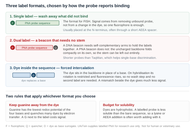
In a hybridisation assay with a wash step, the probe does not need to report anything. Unbound probe is removed, and whatever fluoresces afterwards is bound. A single fluorophore at the N-terminus, usually through a short AEEA spacer to hold the dye off the first base, is the whole requirement. This is the ordinary format for telomere, centromere and bacterial identification probes.
The practical decisions here are about the dye, not the design: whether it survives your fixation and mounting, whether it matches the filter sets on the microscope you will actually use, and whether it bleaches faster than you can acquire. None of those are PNA questions.
A conventional DNA molecular beacon carries a fluorophore at one end and a quencher at the other, with short self-complementary arms that form a stem and hold the two labels together until the probe finds its target. Designing those arms is a real part of the work: too weak and the background is high, too strong and the probe will not open.
A PNA beacon does not need them. The stem-loop structure is not required for quenching in a PNA beacon1; the explanation offered is that the hydrophobic, uncharged PNA backbone tends to adopt a compact structure that minimises exposed hydrophobic surface, which brings a fluorophore and a quencher attached at opposite ends close enough to interact without any sequence designed to do it2. Stemless linear PNA beacons were developed on that basis2.
This removes a design constraint and shortens the probe. In a real-time PCR assay genotyping a single nucleotide polymorphism, PNA beacons labelled with fluorescein or JOE and quenched with QSY-7 were used for allelic discrimination, and the authors note that PNA beacons are significantly shorter than TaqMan or LightCycler probes, which makes both probe design and genotype discrimination easier1. Shorter probes discriminate single-base mismatches better, which is the point of the exercise.
| Common fluorophores | fluorescein/FAM, JOE, TAMRA, Cy3, Cy5, Alexa dyes |
| Common quenchers | dabcyl, QSY-7 and related dark quenchers |
| Placement | fluorophore and quencher at opposite termini; no stem sequence required |
| Spacer | AEEA between dye and first base is usual |
A third option dispenses with the second label. Rather than tethering the dye at a terminus, it is built into the backbone in place of a base. In forced intercalation probes, thiazole orange replaces a base through a short rigid linker; on hybridisation it intercalates at a defined site, its rotation is restricted, and fluorescence rises2. The response is sequence-dependent in a specific and useful way: the largest enhancements occur when an adenine sits next to the dye, and a mismatch adjacent to the dye gives a much smaller signal2.
The alternative single-label design, a light-up probe with thiazole orange tethered at the N-terminus through a flexible linker, has a known weakness worth stating: a significant fraction of the dye binds back onto the probe itself, so performance varies with sequence2. Adding a second dye to a forced-intercalation probe lowers background by more than 99.9%, and a thiazole orange/ITCC FRET pair has given up to a 450-fold enhancement of the acceptor signal2.
This rule is easy to apply at design time and expensive to discover afterwards. Guanine is the most readily oxidised of the four bases, and it quenches many common fluorophores by photoinduced electron transfer. A systematic survey of fluorescein-labelled DNA covering all possible five-base contexts found the expected large loss of fluorescence with guanine proximity, with a terminal thymine giving the strongest signal in labelled single strands and the order running T > A ≈ C ≫ G3. In double-stranded DNA the preference shifts, with a terminal cytosine strongest and the order C ≫ T > A ≫ G, so the terminal G·C pair largely controls emission depending on which of the two the dye sits on3. That study also reports that the change from single- to double-stranded DNA reduces fluorescence intensity by roughly a third3.
That work was done on DNA rather than PNA, and the quantitative ordering should not be transferred to a PNA probe without checking. The underlying interaction is between the dye and the adjacent nucleobase, so the qualitative rule carries: if your sequence allows any choice about where the label goes, do not put it next to a G.
Two consequences are easy to overlook.
The first is solubility. Fluorophores are hydrophobic, and PNA is already prone to aggregation; a labelled probe is less soluble than the same sequence unlabelled. Adding a lysine or an AEEA unit alongside the label is a standard remedy, and solubility enhancers for PNA have been described specifically for this purpose4. We review the labelled sequence rather than the bare one before synthesis for exactly this reason.
The second applies if the probe is going into live cells. A dye is not a passive reporter there. Tetramethylrhodamine- and Alexa Fluor 555-conjugated octaarginine PNAs showed more than a ten-fold increase in antisense activity on irradiation, with the effect correlating with chromophore lipophilicity5. The label is part of the construct's behaviour, not only its readout — which matters for where the conjugate ends up.
If your probe is for PCR rather than microscopy, note that labelled PNA has also been used as a self-reporting element in primers for PCR analysis6, which is a different architecture from a beacon added to the reaction. Tell us which role the probe plays and we will advise on placement.
Send the probe sequence, the dye and quencher you want, which terminus each goes on, and the instrument or filter set the probe has to work with. If you have not chosen dyes, tell us the assay and the available channels and we will suggest a pair.
We review the sequence with the label in place for solubility and flag a guanine sitting next to the planned label position before synthesis rather than after. Labelled probes are purified by HPLC and confirmed by mass spectrometry, so you receive a single labelled species rather than a mixture of labelled and unlabelled material.
Sequences supplied by customers are treated as confidential and are never disclosed or reused.
All LifeTein products are supplied for laboratory research use only. They are not for human or veterinary use, and not for diagnostic or therapeutic use.