


A carrier peptide gets the PNA into the cell. It does not get the PNA out of the compartment it arrives in. Most of a CPP–PNA conjugate taken up by endocytosis stays in endosomes and is eventually degraded, and only the fraction that crosses into the cytosol does anything at all. This is the reason a conjugate that binds its target perfectly can show no effect in cells, and it is a distinct problem from uptake with its own distinct remedies.
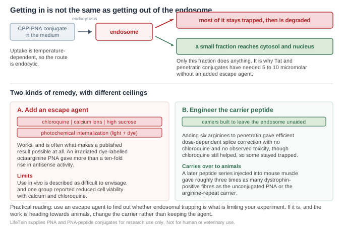
The diagnosis is well established and rests on two observations made together. Uptake of a PNA–oligolysine conjugate was temperature-dependent and led to sequestration of the conjugate in cytoplasmic vesicles, consistent with an endocytic mechanism of internalisation; significant and sequence-specific splicing correction was then achieved only in the presence of endosome-disrupting agents such as chloroquine or 0.5 M sucrose1.
The practical consequence shows up as a concentration requirement. Splicing correction by PNA or morpholino oligomers conjugated to standard carriers such as Tat or penetratin has required 5–10 µM of conjugate unless an endosomolytic agent was added to increase escape from endocytic vesicles2. If your conjugate only works in the micromolar range, endosomal sequestration is the first thing to suspect rather than the binding.
Three treatments are used routinely, and protocols for all three — photodynamic treatment (photochemical internalization), Ca2+ treatment and chloroquine treatment — have been published together specifically for potentiating the antisense effect of CPP–PNA conjugates by increasing release into the cytoplasm3. High sucrose is a fourth1.
Photochemical internalization is the most striking of them. Tetramethylrhodamine- and, to a lesser extent, Alexa Fluor 555-conjugated octaarginine PNAs showed more than a ten-fold increase in antisense activity in the HeLa pLuc705 splice correction assay on irradiation, delivering to the cytosol about as effectively as chloroquine-assisted delivery4. It also makes a point worth remembering when choosing a label: the fluorophore on a conjugate is not necessarily inert, and a lipophilic chromophore can change where the construct ends up.
The limitation is not efficacy. It is that the use of such agents in vivo is difficult to envisage2, which means an experiment built on them does not transfer. There is also a tolerability caveat worth knowing before designing around them: one group reported that in their hands the improvements from calcium and chloroquine were relatively small and that the additions reduced cell viability5.
| Chloroquine | raises endosomal pH; the standard diagnostic for whether trapping is the limit |
| Calcium ions | promotes endosomal rupture; published as a protocol alongside chloroquine |
| High sucrose | 0.5 M has been used to achieve sequence-specific splicing correction |
| Photochemical internalization | light plus a photosensitiser or lipophilic dye; the largest reported effect |
| All four | useful in culture; not a route to an animal experiment |
If adding an agent is the diagnostic, improving the peptide is the fix. Adding six arginines to the N-terminus of penetratin gave a conjugate that produced efficient dose-dependent splice correction in the absence of chloroquine, under conditions where no toxicity was observed2. The same work is honest about the remaining gap: adding chloroquine still improved activity, which shows that some of the conjugate was still entrapped2. Better carriers reduce the problem rather than abolishing it.
The reason this is the right direction is that it survives the move out of culture. A later series of PNA internalisation peptides was tested in animals: injected into the tibialis anterior muscles of mdx mice, the conjugates produced roughly three times as many dystrophin-positive fibres as either the unconjugated PNA or the conjugate carrying the arginine-repeat carrier6. No endosomolytic agent is involved in that result.
Which carrier to attach in the first place, how the linkage and the peptide's position affect activity, and the published head-to-head comparisons are covered in CPP–PNA conjugate design.
Send the PNA sequence, the carrier peptide and which terminus it should sit on, and tell us whether the work will stay in culture or move to an animal model — that distinction changes which carrier is worth using. If you intend to label the conjugate, say so at the quoting stage rather than afterwards, since the label is part of the construct and affects both the synthesis route and the behaviour described above.
We build the peptide and the PNA as a single construct where the chemistry allows, review the combined sequence for solubility before synthesis, and include purification and mass confirmation.
Sequences supplied by customers are treated as confidential and are never disclosed or reused.
All LifeTein products are supplied for laboratory research use only. They are not for human or veterinary use, and not for diagnostic or therapeutic use.