


Binding a single-stranded target is the easy case. Binding double-stranded DNA means competing with a strand that is already there, and the DNA duplex does not give up its partner willingly. PNA is one of the few synthetic oligomers that can win that competition, which is what makes antigene work — acting on the gene rather than the message — possible at all. There are three established ways to do it, and the right question is not which is best but which one your target sequence and your buffer will allow.
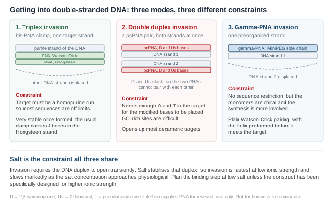
PNA has no phosphate backbone, so a PNA–DNA hybrid carries none of the charge repulsion that destabilises a DNA–DNA duplex. The hybrid is therefore more stable than the DNA duplex it replaces, and that difference in stability is the energy that drives strand displacement. The original demonstration used a thymine-substituted polyamide against a double-stranded target and showed sequence-selective recognition by strand displacement rather than by binding in a groove1.
The reaction still has to wait for an opportunity. A DNA duplex opens transiently, and the PNA can only enter during one of those openings. Anything that stabilises the duplex — higher salt above all — reduces how often those openings occur and slows invasion accordingly. This is the single most common reason a duplex-invasion experiment that worked in one buffer fails in another.
The classical approach uses a homopyrimidine PNA that binds the purine strand of the target twice: once by Watson–Crick pairing and once, folded back, by Hoogsteen pairing. The displaced DNA strand is left as a single-stranded loop, a structure usually called a P-loop. The resulting complex is extremely stable, which is why this mode remains useful despite its limitation.
The limitation is severe and it is sequence, not chemistry: the target must be a homopurine run. Most genomic sites are not. The Hoogsteen strand of such a clamp conventionally carries pseudoisocytosine in place of cytosine so that it does not depend on the acidic pH that protonated cytosine would require; that substitution is covered separately in pseudoisocytosine and PNA triplex stability.
The second approach removes the homopurine restriction by changing the bases rather than the backbone. Two PNA oligomers, each complementary to one of the two DNA strands, are synthesised with adenine replaced by 2,6-diaminopurine (D) and thymine replaced by 2-thiouracil (Us). D pairs normally with T and Us pairs normally with A, so each PNA binds its DNA strand well. But D and Us clash sterically with each other, so the two PNAs cannot form a PNA–PNA duplex and sequester each other2.
The result is that both DNA strands can be targeted simultaneously, each by one PNA, with the duplex unwound between them. The authors of the original work predicted that for decameric targets more than 80% of all sequences could be addressed this way by straightforward Watson–Crick pairing, and demonstrated the consequence directly: pseudo-complementary PNAs directed at the T7 RNA polymerase promoter inhibited transcription initiation2. That is antigene activity in the literal sense — the gene is blocked at the DNA level, not the transcript.
The residual constraint is composition. The modified bases replace A and T, so a target with very little A or T offers few positions to modify and invasion becomes inefficient. Adding positive charge to the backbone addresses this: when the pseudo-complementary bases were carried on chiral units derived from N-(2-aminoethyl)-d-lysine, invasion succeeded even at strongly GC-rich sites such as (G/C)7(A/T)3 and (G/C)8(A/T)2, which were otherwise hardly targetable3.
| D | 2,6-diaminopurine — replaces adenine; pairs with T, clashes with Us |
| Us | 2-thiouracil — replaces thymine; pairs with A, clashes with D |
| Ordered as | a matched pair of PNAs, one per DNA strand, with the substitutions at every A and T position |
| Target composition | works best with reasonable A/T content; GC-rich targets need backbone modification |
The third approach leaves the bases alone and preorganises the backbone. Installing a small hydrophilic (R)-diethylene glycol group — MiniPEG — at the gamma position of each PNA unit turns a randomly folded oligomer into a right-handed helix before it ever meets its target. Oligomers made this way hybridise with greater affinity and sequence selectivity than the unmodified parent, and are more water-soluble and less prone to aggregate4.
Because the entropic cost of folding has already been paid, a single such strand carries enough binding free energy to invade a double helix on its own, without a clamp and without modified bases. Sequence-unrestricted recognition of double-helical B-DNA by (R)-MiniPEG gamma-PNAs was demonstrated by gel-shift assay5. This is the most general of the three modes and also the most demanding to synthesise, since every monomer is chiral.
Salt remains the boundary. With ordinary nucleobases, gamma-PNA invasion is confined to sub-physiological ionic strength; recognition under physiological conditions has been achieved by raising the binding free energy further, for instance by incorporating G-clamp nucleobases6. The practical reading for a laboratory is that an invasion step should be planned at low salt unless the construct has been designed specifically to work above it.
| Mode | What you order | Target requirement | Typical use |
|---|---|---|---|
| Triplex invasion | one bis-PNA clamp, usually with J bases in the Hoogsteen arm | homopurine stretch | very stable complexes at a permissive site; PCR clamping chemistry |
| Double duplex invasion | two pseudo-complementary PNAs, D and Us substituted | mixed sequence with adequate A/T | blocking a promoter or other defined duplex site |
| Gamma-PNA invasion | one gamma-modified PNA, MiniPEG side chains | none in principle | mixed sequences where the other two modes do not apply |
Antigene work is a harder proposition than antisense for a reason that has nothing to do with binding: the target is in the nucleus, inside chromatin, and present at two copies per cell rather than at the thousands of copies a transcript provides. Binding chemistry that works cleanly on a plasmid in a tube is only the first requirement. Delivery is the practical obstacle, as it is for antisense PNA, and nuclear access adds a further step on top of it.
For a duplex-invasion construct, send the double-stranded target sequence rather than a single strand, and say which mode you intend. If you are not sure, send the sequence and the buffer you plan to bind in — the composition usually settles the question. For a pseudo-complementary pair we synthesise both oligomers together so the substitution pattern is matched across the two. For gamma-PNA we will confirm the modification pattern before synthesis, since placing MiniPEG at every position and at selected positions give different products.
We will also review the sequence for solubility before synthesis and suggest a lysine or AEEA addition where the composition calls for one. Purification and mass confirmation are included.
Sequences supplied by customers are treated as confidential and are never disclosed or reused.
All LifeTein products are supplied for laboratory research use only. They are not for human or veterinary use, and not for diagnostic or therapeutic use.