


Most of the difficulty people have with PNA happens in the first hour after the tube arrives, and almost none of it is the sequence's fault. PNA has no charged backbone, which is the property that makes it bind so tightly — and the same property makes it less willing to dissolve than the equivalent DNA oligonucleotide, and makes its concentration easier to get wrong. Both problems are avoidable if you know they are coming.
The original design replaced the sugar–phosphate backbone with a neutral polyamide1. A DNA oligonucleotide carries one negative charge per residue and is pulled into solution by them; PNA has none. The molecules are therefore free to stack on one another, and the same base-stacking that gives PNA its affinity for a target also drives self-aggregation.
Composition decides how badly. Published commercial design guidelines converge on the same factors — high purine content, long runs of consecutive purines, high G content and greater length all reduce aqueous solubility — though the specific thresholds differ between suppliers, so treat them as directional rather than absolute. Self-complementary stretches are a separate hazard, because PNA–PNA pairing is stronger than PNA–DNA pairing, so a probe that can bind itself will.
If the composition is unavoidable, the remedy is to build solubility in rather than fight for it afterwards. Attaching one or two lysine residues or a short AEEA linker is standard practice, and solubility enhancers for PNA have been described in the literature for exactly this purpose2. It is far cheaper to add a lysine at the design stage than to discover the problem with the material in hand.
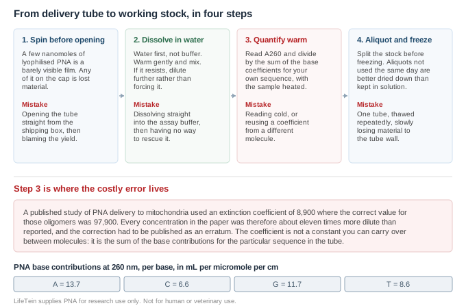
Work in water, not buffer. Purified PNA arrives from reversed-phase HPLC with trifluoroacetic acid, which leaves the basic amino groups protonated — and that residual charge is part of what keeps the material in solution at stock concentrations. Dissolving straight into a neutral assay buffer discards that advantage at the worst possible moment, and leaves you with nothing to adjust.
The sequence to work through:
If the material is labelled, be more cautious with heat. A fluorophore is the least thermally robust part of the construct, and the label is also what made the probe less soluble in the first place.
This is the step worth slowing down for, because an error here is invisible — everything still works, it just works at a different concentration than you recorded.
The cautionary case is a published one. In a study of PNA delivery to human mitochondria, concentrations were calculated using an extinction coefficient of 8,900 M−1cm−1 when the correct value for those oligomers was 97,900 M−1cm−1; as the published erratum states, every concentration in the paper was consequently about eleven-fold more dilute than reported3. That is not a careless laboratory — it is a coefficient being treated as a property of the molecule class rather than of the specific sequence.
The coefficient is the sum of the base contributions for the sequence in your tube. The per-base values in common use for PNA are listed below4; add them up for your own sequence, multiply by nothing else, and divide the absorbance by the result.
| Base | ε260 per base (mL µmol−1 cm−1) |
|---|---|
| A | 13.7 |
| C | 6.6 |
| G | 11.7 |
| T | 8.6 |
Measure the sample warm. Residual structure suppresses absorbance, so a room-temperature reading understates the true value: coefficients determined from room-temperature measurements have been reported to run 5–10% lower than those measured at elevated temperature, and warming the sample before reading is the standard remedy5. Repeated heating does not harm PNA, so there is no reason to avoid it.
Two things the A260 figure does not tell you. It does not distinguish PNA from anything else in the tube that absorbs at 260 nm, and it says nothing about how much of the dry mass is peptide rather than counterion and water. If you need the second number, net peptide content is available as a separately quoted test with its own fee rather than something measured on every order — ask for it at the quoting stage if your work depends on accurate mass. Counterion exchange is likewise quoted separately.
Lyophilised PNA is the stable form, and PNA itself is resistant to both nucleases and proteases, so biological degradation is not what you are guarding against. What you are guarding against is loss of material to tube walls, and loss of soluble material to slow aggregation.
Two pieces of data come with the material, and they answer different questions.
| Method | What it establishes | What it does not |
|---|---|---|
| Reversed-phase HPLC | How much of the material is the main species rather than deletion sequences and other synthesis by-products | Whether the main species is the right molecule |
| Mass spectrometry | That the main species has the expected mass, which confirms the sequence length, the modification and the label are present | The purity, or the position of an internal modification |
You need both, and they are supplied together. A clean chromatogram with the wrong mass means a well-purified wrong product; the right mass with a poor chromatogram means the molecule is correct but accompanied. For a dual-labelled probe the mass is also the practical check that both labels survived synthesis and cleavage, which is not something the chromatogram will tell you on its own.
We review the sequence for solubility before synthesis rather than after, and will propose a lysine or AEEA addition where the composition calls for one — with the label included in that assessment, not just the bases. Material is purified by reversed-phase HPLC and confirmed by mass spectrometry, and both are supplied with the order.
If a sequence looks likely to be difficult, we would rather raise it at the quoting stage than deliver material you cannot get into solution. Tell us the buffer and the assay you intend to use and we will flag anything that is going to fight you.
Sequences supplied by customers are treated as confidential and are never disclosed or reused.
All LifeTein products are supplied for laboratory research use only. They are not for human or veterinary use, and not for diagnostic or therapeutic use.