


The single most common mistake in PNA FISH is running it like DNA FISH. The conditions are not a minor variation on the same protocol — the salt concentration moves in the opposite direction, and for a reason that follows directly from the chemistry. This page explains why, what each buffer component is doing, and how to tell an accessibility problem from a stringency problem when the slide comes out wrong.
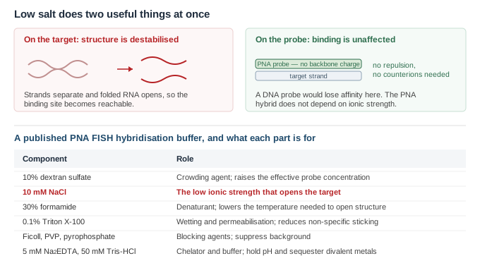
In conventional hybridisation, salt is required. Both probe and target carry a negatively charged phosphate backbone, they repel one another, and cations are needed to screen that repulsion before a duplex can form. Lowering the salt in a DNA FISH experiment weakens the probe.
PNA has no backbone charge, so there is nothing to screen1. Hybridisation is less sensitive to salt concentration for exactly that reason, and the absence of electrostatic repulsion also raises the hybridisation rate relative to DNA probes2. This leaves the ionic strength free to be used for something else — and what it is used for is the target.
Stated directly in the published method: the uncharged backbone allows hybridisation to be performed under low-salt conditions that are denaturing for ribosomal RNA secondary structure but favourable for PNA–RNA hybridisation3. Low salt destabilises the target's own folding and the reannealing of complementary chromosomal strands, while leaving the PNA hybrid intact. The two effects point the same way, which is what makes the method work on folded RNA and on double-stranded chromosomal targets alike.
A published PNA FISH hybridisation buffer contains 10% dextran sulfate, 10 mM NaCl, 30% formamide, 0.1% sodium pyrophosphate, 0.2% polyvinylpyrrolidone, 0.2% ficoll, 5 mM Na2EDTA, 0.1% Triton X-100 and 50 mM Tris-HCl3. Each component has a job.
| Component | What it does | If you change it |
|---|---|---|
| 10 mM NaCl | Sets the low ionic strength that destabilises target structure | Raising it re-folds the target and can abolish the signal; this is the component not to borrow from a DNA protocol |
| 30% formamide | Denaturant; lowers the temperature at which structure opens | More formamide raises stringency and lowers signal; less increases background |
| 10% dextran sulfate | Crowding agent; raises the effective probe concentration | Omitting it slows hybridisation and usually costs signal |
| 0.1% Triton X-100 | Wetting and permeabilisation; reduces non-specific adsorption | Omitting it commonly shows up as patchy, uneven staining |
| Ficoll, PVP, pyrophosphate | Blocking agents that suppress background | Omitting them raises background without affecting specific signal |
| Na2EDTA, Tris-HCl | Chelator and buffer | Routine; hold pH and sequester divalent metals |
The hybridisation step decides how much probe binds; the wash decides how much of it was bound correctly. For PNA this matters more than usual, because the mismatch discrimination the probe is capable of is only realised if the wash is stringent enough to exploit it.
That discrimination is real but not uniform. Against an 11-mer PNA, single-base mismatches cost more than 20 °C of melting temperature at the least tolerated positions, and the size of the penalty depended on both where the mismatch sat and which bases were paired: G:T and T:T were the most tolerated, A:A and G:G the most destabilising4. A wash set for the worst case will strip a tolerated mismatch and leave the specific signal; a wash set too gently leaves both.
Change one variable at a time, and change the wash before the hybridisation. Wash temperature is the cleanest single lever: it acts only on what is already bound, so it separates specific from non-specific binding without altering how much probe reached the target in the first place. Adjusting formamide or salt changes both at once, which is why a protocol tuned by moving several variables together rarely transfers to the next probe.
These are different problems with almost no overlap in their causes, and the first step is deciding which one you have. A universal probe in a second colour settles it immediately: if the universal signal is also absent, nothing reached the target and the problem is upstream of stringency.
| No signal anywhere | Fixation or permeabilisation; probe never reached the target; salt too high so the target stayed folded; dead fluorophore |
| No specific signal, universal probe fine | Target region inaccessible within the folded structure, or the probe sequence is wrong for the organism present |
| Signal everywhere | Wash too gentle; probe concentration too high; blocking agents omitted; autofluorescence of the matrix rather than probe at all |
| Patchy or uneven | Detergent omitted; smear too thick; incomplete coverage during hybridisation |
| Signal fades during imaging | Photobleaching; use an antifade mountant and set exposure on a sacrificial field |
Autofluorescence deserves separate mention because it is routinely mistaken for background. It is not removed by any wash, since nothing is bound. If raising stringency does not reduce it, image an unhybridised control: whatever is still glowing is the matrix, and the answer is a red or far-red fluorophore rather than a harsher protocol.
Solubility is a design question as much as a handling one, and is covered separately in the PNA Knowledge Center.
Send us the target sequence or the organism and region you need to discriminate, plus the fluorophore your filter set is built around. We supply the PNA purified with the analytical data for the batch. Hybridisation and wash conditions have to be established on your own material, since accessibility and autofluorescence depend on the sample rather than on the probe.
Sequences supplied by customers are treated as confidential and are never disclosed or reused.
All LifeTein products are supplied for laboratory research use only. They are not for human or veterinary use, and not for diagnostic or therapeutic use.