


Most molecular identification methods begin by destroying the sample: lyse the cells, extract the nucleic acid, amplify it. PNA FISH does none of that. A labelled probe is hybridised to ribosomal RNA inside intact, fixed cells on a slide, and the organism is identified by looking at it. The method works because ribosomal RNA is abundant, because its sequence carries species-level information, and because a PNA probe can cross a fixed cell envelope and bind a structured RNA target that a DNA probe struggles with.
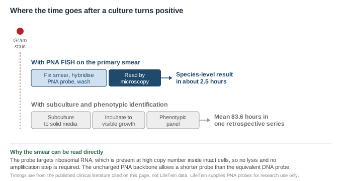
Three properties make rRNA an unusually good in-situ target. It is present at very high copy number, because a growing bacterium carries thousands of ribosomes, so the signal from a single cell is strong enough to see without any amplification step. Its sequence contains regions conserved across all bacteria interleaved with regions that vary between species, so a probe can be designed to be as broad or as narrow as the question requires. And it is a natural cellular component rather than a single-copy gene, so detection does not depend on the cell having been lysed efficiently.
The cost of that abundance is structure. Ribosomal RNA is extensively folded and bound by protein, and a probe has to displace or thread into that structure to reach its target. This is where the chemistry matters.
PNA binds complementary nucleic acid following Watson-Crick rules but with no phosphate charge on its own backbone, which removes the electrostatic repulsion that limits how tightly a DNA probe can bind1. Two practical consequences follow for FISH.
The first is that a PNA probe can be shorter for the same specificity. In the work that established the method for Staphylococcus aureus, the PNA probe was designed against the same species-specific region of 16S rRNA that earlier DNA probes had targeted, but was made shorter than those DNA probes precisely because its affinity was higher2. A shorter probe diffuses into fixed cells more readily and is less likely to be trapped by secondary structure.
The second is that hybridisation can be run at low ionic strength. A published PNA FISH hybridisation buffer contains 10 mM NaCl3 — an order of magnitude or more below what a DNA probe would need — because the uncharged backbone does not require counterions to approach its target. Low salt destabilises the RNA's own internal structure while leaving the PNA hybrid intact, which is what opens the target up.
| Target | 16S rRNA (bacteria) or 18S/26S rRNA (yeast), inside fixed intact cells |
| Sample | A smear, made directly from culture; no extraction, no amplification |
| Probe length | Typically around 15 bases, shorter than the equivalent DNA probe2 |
| Hybridisation | Low-salt, formamide-containing buffer; see the published protocol3 |
| Read-out | Epifluorescence microscopy, with cell morphology visible alongside the signal |
The method was characterised most thoroughly for S. aureus in positive blood cultures. An initial evaluation against 17 reference strains and 48 clinical isolates, including methicillin-resistant and methicillin-susceptible S. aureus, coagulase-negative staphylococci and related organisms, reported 100% sensitivity and 96% specificity, with a result available in about 2.5 hours2. A subsequent blinded multicentre comparison across eight clinical microbiology laboratories and 564 blood culture bottles reported sensitivity and specificity of 100% and 99.2% in one bottle system and 98.5% and 98.5% in another4.
The approach was later extended beyond Gram-positive cocci. The first application to Gram-negative rods reported correct identification of 32 of 34 blood cultures growing Pseudomonas aeruginosa5. A retrospective clinical series covering both bacteria and Candida reported identification by PNA FISH at a mean of 11.2 hours against a mean of 83.6 hours for conventional culture, with identification accuracy for bacteria of 100% in 73 of 73 blood cultures6.
Those figures come from clinical studies of validated diagnostic assays, and they describe what the chemistry is capable of in trained hands on a defined sample type. They are not performance claims for a research probe in a new application. A probe designed against a different organism, used on a different matrix, has to be characterised on that matrix.
Probe length, fluorophore, and the hybridisation and wash conditions interact, and the usual approach is to fix the probe and tune the wash. The PNA Knowledge Center indexes the related design pages as they are published.
Send us the target sequence, or the organism and the region you want to discriminate, together with the fluorophore your filter set is built around. We synthesise and label the PNA and supply it purified, with the analytical data for the batch. We do not validate probes against clinical specimens and we do not supply diagnostic assays.
Sequences supplied by customers are treated as confidential and are never disclosed or reused.
All LifeTein products are supplied for laboratory research use only. They are not for human or veterinary use, and not for diagnostic or therapeutic use. The clinical studies cited on this page describe separately validated diagnostic assays and are included as scientific background; they are not claims about LifeTein products.