


A centromeric probe answers a different question from a telomeric one. Telomere work measures how much signal is present; enumeration asks only how many discrete signals there are. That makes the assay conceptually simple and the failure modes entirely about scoring. This page covers why chromosome-specific centromeric probes are possible at all, what PNA contributes, and where counts go wrong.
Human centromeres sit in arrays of alpha satellite, a repeat built on a monomer of about 171 base pairs. Individual monomers are only 50 to 70% identical to one another, and a defined number of them are arranged in tandem to form a higher-order repeat unit. It is the number of monomers and their order within that higher-order unit that confers chromosome specificity, so the centromeres of different chromosomes are defined by structurally distinct higher-order repeats1.
Those higher-order units are then reiterated hundreds to thousands of times, producing arrays that are 97 to 100% internally homogeneous and extend over megabases1. Two consequences follow for probe design. The target is enormous, which is why a centromeric signal is bright enough to see easily. And the discrimination a probe needs is between closely related repeat families rather than between unrelated sequences, which is where probe chemistry starts to matter.
PNA binds complementary nucleic acid by Watson-Crick pairing but carries no charge on its backbone, so probe and target do not repel one another and binding is tighter than the equivalent DNA duplex2. For a repetitive target this has a practical effect: the probe competes successfully against reannealing of the chromosomal strands themselves, which is the main obstacle to in-situ hybridisation at a tandem repeat.
Discrimination between near-identical repeat families is the other half. In a systematic study of an 11-mer PNA against DNA carrying every single-base mismatch at every position, the melting temperature penalty exceeded 20 °C for the least tolerated placements and depended on both the position of the mismatch and the identity of the bases involved3. That sensitivity is what allows a short probe to separate one alpha-satellite family from another.
In practical terms the result is a bright centromeric signal in metaphase spreads and discrete fluorescent spots in interphase nuclei4.
| Target | Chromosome-specific alpha-satellite higher-order repeat array |
| Read-out | Number of discrete signals per nucleus |
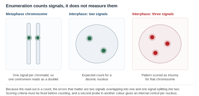
| Metaphase appearance | A doublet at one centromere, one signal per sister chromatid |
| Interphase appearance | One compact spot per copy of the chromosome |
| Typical use | Copy-number screening where banding is impractical or no dividing cells are available |
Centromeric PNA probes have been used for rapid in-situ identification of human chromosomes and for detecting aneuploidy, and multicolour protocols identifying several chromosomes at once have been described5. Reported applications span lymphocytes, amniocytes and spermatozoa as well as isolated oocytes and blastomeres5, and the approach has been applied specifically to human oocytes, polar bodies and preimplantation embryos6. The chemistry and its cytogenetic rationale are reviewed in more detail elsewhere7.
Those studies are published research applications, several of them in a reproductive-genetics setting. LifeTein supplies labelled PNA probes for research use. We do not supply diagnostic assays, and we do not validate probes against clinical specimens.
Because the read-out is a number rather than an intensity, the usual sources of error in quantitative FISH do not apply. Two different ones do, and both are scoring problems rather than chemistry problems.
| Artefact | What it looks like | What it does to the count |
|---|---|---|
| Overlapping signals | Two homologues lying close together in the nucleus, read as one spot | Undercount; a disomic nucleus scored as monosomic |
| Split signals | One array resolved into two closely spaced spots, often in a decondensed nucleus | Overcount; a disomic nucleus scored as trisomic |
| Overlapping nuclei | Two nuclei not separated in the image | Apparent high copy number |
| Truncated nuclei | Part of a nucleus lost in sectioning or at the field edge | Undercount |
Tell us which chromosome you need to target and the fluorophore your filter set is built around. If you are running a two-colour enumeration we can supply the test and reference probes from the same synthesis with spectrally separated labels, which removes one source of batch-to-batch variation between the two channels.
Sequences supplied by customers are treated as confidential and are never disclosed or reused.
All LifeTein products are supplied for laboratory research use only. They are not for human or veterinary use, and not for diagnostic or therapeutic use.