
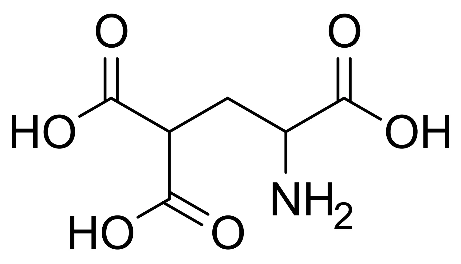
Gamma-Carboxyglutamic acid (GLA) is a unique non-proteinogenic amino acid that plays an indispensable role in a select group of proteins, most notably those involved in blood coagulation and calcium-dependent biological processes. Unlike standard amino acids, GLA is not directly encoded by DNA; instead, it is generated through a vitamin K-dependent post-translational modification of glutamic acid residues. This modification introduces a second carboxyl group onto the γ-carbon of the glutamate side chain, transforming a simple amino acid into a potent calcium-binding motif. This unique structural feature is critical for the function of coagulation factors, bone proteins, and certain neurotoxins, making GLA a molecule of significant interest in both basic research and therapeutic development.
Key Takeaways
- Gamma-Carboxyglutamic acid (GLA) is a non-proteinogenic amino acid formed by the vitamin K-dependent carboxylation of glutamic acid residues.
- Its dicarboxylic acid side chain confers unique metal-binding properties, particularly for calcium ions (Ca²⁺).
- GLA is essential for the biological activity of vitamin K-dependent coagulation factors (e.g., prothrombin, Factor VII, Factor IX, Factor X) and bone proteins such as osteocalcin.
- The synthesis of GLA-containing peptides is challenging due to the lability of the malonic acid-like side chain, which is prone to decarboxylation under acidic conditions.
- Successful solid-phase peptide synthesis (SPPS) relies on the use of orthogonally protected building blocks, most commonly Fmoc-L-Gla(OtBu)₂-OH, to prevent side reactions and ensure correct incorporation.
- Specialized providers like LifeTein offer custom synthesis of GLA-containing peptides, enabling researchers to study calcium-dependent protein interactions and develop novel therapeutics.
Chemical Fundamentals of Gamma-Carboxyglutamic Acid
Structure and Uniqueness
Gamma-carboxyglutamic acid is defined by a dicarboxylic acid side chain attached to the α-carbon. This structure differs from glutamic acid by the addition of a second carboxyl group at the γ-position, creating a malonic acid-like motif. This unique metal-binding property allows GLA residues to chelate calcium ions, conferring metal-binding character to the proteins into which they are incorporated. The presence of two carboxylic acid groups gives GLA a strong negative charge at physiological pH, enabling it to interact with positively charged surfaces, such as phospholipid membranes.
Biosynthesis: A Vitamin K-Dependent Modification
GLA is synthesized through the post-translational carboxylation of specific glutamic acid residues within a protein sequence. This reaction is catalyzed by vitamin K-dependent carboxylase and requires reduced vitamin K, oxygen, and carbon dioxide. This modification is not encoded by the genetic code; instead, it is a dynamic process that allows organisms to fine-tune the calcium-binding properties of specific proteins in response to physiological needs.
Find out more about peptide synthesis here.
Biological Significance and Function
Role in Blood Coagulation
GLA is most renowned for its essential role in the blood coagulation cascade. It is a key component of several vitamin K-dependent coagulation factors, including prothrombin (Factor II), Factor VII, Factor IX, and Factor X, as well as the regulatory proteins Protein C, Protein S, and Protein Z. The GLA residues in these proteins function as ligands for Ca²⁺, which are crucial for their biological activity. Calcium binding induces conformational changes that allow these factors to assemble on the surface of activated platelets, a critical step for effective clot formation.
Function in Bone and Other Tissues
Beyond coagulation, GLA is found in proteins of calcified tissues, most notably osteocalcin (bone-Gla protein). Osteocalcin contains multiple GLA residues that enable it to bind to hydroxyapatite crystals in bone, playing a role in bone mineralization and turnover. GLA has also been observed in renal calculi, atherosclerotic plaque, and the egg chorioallantoic membrane, suggesting diverse physiological and pathological roles.
Occurrence in Natural Toxins
GLA is also present in certain neurotoxic peptides from animal venoms. For example, conantokin-G, isolated from the venom of the marine snail Conus geographus, contains GLA residues that are critical for its activity as an NMDA receptor antagonist. This highlights the broad evolutionary conservation of GLA as a calcium-binding module.
Synthesis of GLA-Containing Peptides
The Challenge of Chemical Synthesis
The synthesis of peptides containing GLA is notoriously difficult. The primary challenge stems from the malonic acid-like structure of the GLA side chain, which is susceptible to decarboxylation, particularly under the acidic conditions commonly used in peptide synthesis and cleavage. Furthermore, the presence of two carboxylic acid groups necessitates a robust orthogonal protection strategy to prevent unwanted side reactions during peptide chain elongation.
Orthogonal Protection Strategies
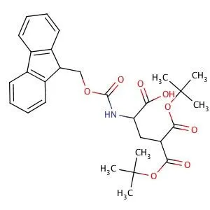
To overcome these challenges, researchers rely on orthogonally protected GLA building blocks. The most widely used and commercially available derivative is Fmoc-L-Gla(OtBu)₂-OH (CAS 308357-14-4). In this building block:
- The α-amino group is protected by the base-labile Fmoc group.
- The two γ-carboxyl groups of the GLA side chain are protected as di-tert-butyl esters (OtBu) .
This orthogonal protection scheme ensures that the Fmoc group can be removed under mild basic conditions (e.g., piperidine) during each synthesis cycle, while the OtBu groups remain stable and are only cleaved by strong acid (e.g., TFA) during the final deprotection step. This strategy prevents the premature decarboxylation and lactonization of the GLA residue, ensuring its integrity throughout peptide assembly.
Improved Synthetic Outcomes
Comparative studies have demonstrated that employing Fmoc-Gla(OtBu)₂-OH as a pre-protected building block significantly improves synthesis outcomes. It has been shown to increase overall peptide yield by 21 percentage points and reduce side product formation compared to attempts to incorporate GLA via post-synthetic carboxylation or using less robustly protected derivatives. This highlights the importance of using fully protected building blocks for successful GLA peptide synthesis.
Find out about high-speed RUSH synthesis.
Frequently Asked Questions (FAQ)
What is the difference between glutamic acid and gamma-carboxyglutamic acid?
Glutamic acid has a single carboxyl group in its side chain. Gamma-carboxyglutamic acid (GLA) has two carboxyl groups attached to the γ-carbon, creating a dicarboxylic acid structure that is essential for calcium binding.
Why is GLA considered an “unusual” amino acid?
GLA is “unusual” because it is not directly encoded by DNA. It is created through a post-translational modification of glutamic acid residues, a process that requires vitamin K.
What is the function of GLA in proteins?
The primary function of GLA is to act as a high-affinity calcium-binding site. This calcium binding is essential for the biological activity of blood coagulation factors and for the function of bone proteins like osteocalcin.
Why is it difficult to synthesize peptides containing GLA?
The synthesis is challenging because the GLA side chain is prone to decarboxylation under acidic conditions. Additionally, its two carboxylic acid groups require a carefully designed orthogonal protection strategy to prevent side reactions during solid-phase peptide synthesis.
What is the standard building block for GLA in peptide synthesis?
The most commonly used building block is Fmoc-L-Gla(OtBu)₂-OH. It features an Fmoc-protected amine and two tert-butyl-protected carboxyl groups, allowing for standard Fmoc/tBu SPPS workflows.
References
Zhang, C.-Y., Wu, W.-H., Wang, J., & Lan, M.-B. (2012). Antioxidant Properties of Polysaccharide from the Brown Seaweed Sargassum graminifolium (Turn.), and Its Effects on Calcium Oxalate Crystallization. Marine Drugs, 10(1), 119–130. https://doi.org/10.3390/md10010119