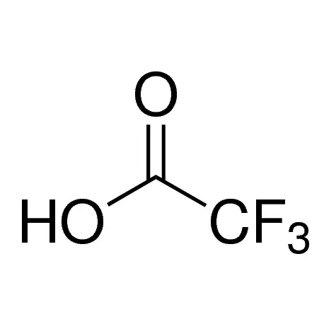
Trifluoroacetic acid (TFA) is ubiquitous in peptide synthesis, serving as a cleavage reagent during solid-phase synthesis and as an ion-pairing agent in HPLC purification. Consequently, synthetic peptides are typically delivered as TFA salts. While TFA facilitates high-purity peptide production, its presence as a salt can profoundly compromise experimental outcomes and biological activity. The decision to remove TFA hinges on your peptide’s intended application, sequence properties, and sensitivity requirements.
Key Takeaways
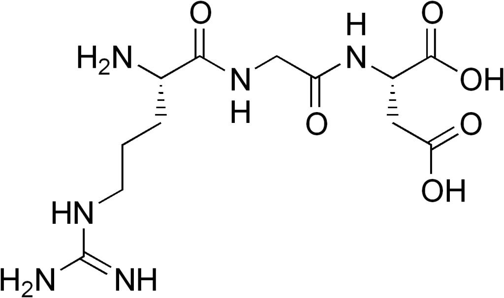
- Residual TFA alters peptide structure and function by binding to positively charged residues, potentially modifying mass, solubility, and secondary structure.
- TFA is cytotoxic at nM concentrations, interfering with cell proliferation, receptor binding, and enzymatic activity in biological assays.
- HCl exchange is the gold-standard removal method, replacing TFA counterions via iterative lyophilization in hydrochloric acid.
- Critical applications like cellular assays, in vivo studies, or API development mandate TFA levels <1%.
- TFA sensitivity varies; hydrophilic peptides or those with cationic residues (Arg, Lys, His) bind TFA more tightly, necessitating aggressive removal.
Biological Assay Interference: A Primary Concern
Cytotoxicity and Cellular Dysregulation
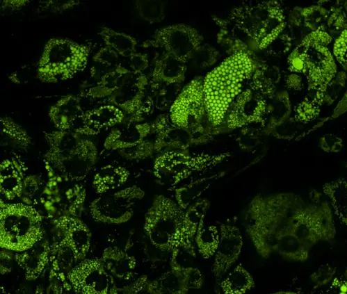
TFA exhibits dose-dependent cytotoxicity, disrupting membrane integrity, inhibiting cell proliferation, and triggering apoptosis at concentrations as low as 10 nM. For cell-based assays—especially those measuring viability, signaling, or metabolism—TFA removal is non-negotiable.
Enzymatic and Receptor Binding Interference
The strong acidity of TFA (pKa 0.23) can denature pH-sensitive proteins or enzymes, leading to false-negative results in kinetic assays. Additionally, TFA competes with phosphate groups in binding sites, potentially inhibiting kinases, phosphatases, or ATP-dependent enzymes. For studies probing enzyme-substrate interactions or receptor-ligand binding, TFA levels should be reduced below 1% using professional exchange services.
Structural and Functional Consequences of TFA Retention
Altered Peptide Conformation and Solubility
TFA binds tightly to free amino termini and side chains of cationic residues (e.g., Arg, Lys, His), forming stable counterion complexes that distort secondary structures like α-helices or β-sheets. This binding can reduce solubility in aqueous buffers and promote aggregation, particularly in hydrophobic sequences. For structural biology applications (e.g., NMR, crystallography), TFA removal ensures native folding and minimizes artifacts.
Applications Dictating TFA Removal
In Vivo Studies and Therapeutic Development
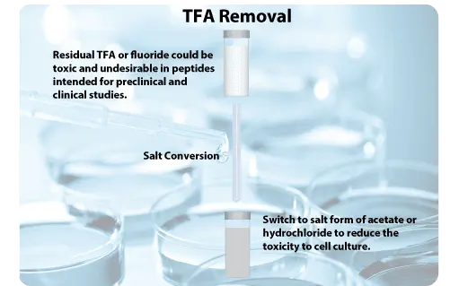
For peptides intended for animal studies or clinical use, TFA poses safety and efficacy risks. Its toxicity profile includes organ toxicity and immunogenicity, potentially invalidating preclinical data. Regulatory guidelines for Active Pharmaceutical Ingredients (APIs) require TFA levels <0.1%, necessitating rigorous removal protocols like LifeTein’s TFA Salt Exchange.
Practical Removal Methodologies
HCl Exchange Protocol
LifeTein’s optimized protocol replaces TFA with HCl through iterative dissolution and lyophilization:
- Dissolve peptide in distilled water (1 mg/mL) or phosphate buffer.
- Add 100 mM HCl to achieve 2–10 mM final concentration.
- Incubate 1 minute at room temperature.
- Flash-freeze in liquid nitrogen.
- Lyophilize overnight, then repeat dissolution in HCl and lyophilization twice.
- Resuspend in target buffer at 2 mg/mL.
Note: Concentrations <2 mM HCl yield incomplete exchange, while >10 mM risks peptide modification.
Professional TFA Exchange Services
For stringent requirements (e.g., <1% TFA), specialized services like LifeTein’s TFA Salt Exchange replace TFA with acetate, formate, or HCl. This approach is recommended for hydrophilic peptides or complex sequences where DIY methods fail.
Decision Workflow: When to Remove TFA
Evaluate your experimental needs using this framework:
- Remove TFA if: Conducting cellular assays, in vivo work, structural studies, or MS quantification.
- Tolerable if: Using peptides for polyclonal antibody production or non-quantitative Western blotting.
Frequently Asked Questions (FAQ)
Can I use acetate instead of HCl for TFA exchange?
Yes. Acetate or formate salts are less acidic alternatives, though HCl offers higher exchange efficiency for strongly cationic peptides.
Does lyophilization alone remove TFA?
No. Lyophilization eliminates unbound TFA but not counterions bound to peptide residues. HCl exchange or HPLC desalting is essential for bound TFA.
Are TFA-free peptides more expensive?
Yes. Salt conversion services incur 20–30% higher costs due to peptide loss during purification and additional reagents.