Unusual Amino Acids: 2,6-Diaminopimelic Acid (DAP)

DAP

2,6-Diaminopimelic acid (DAP) is a unique non-proteinogenic diamino acid that occupies a unique position at the intersection of bacterial physiology, antibiotic development, and synthetic peptide chemistry. Structurally characterized as an α,α′-diamino dicarboxylic acid with the molecular formula C7H14N2O4, DAP is distinguished by the presence of two chiral centers and two amino groups flanking a seven-carbon backbone. This unusual amino acid is an essential constituent of bacterial peptidoglycan and serves as the biosynthetic precursor to L-lysine in both bacteria and higher plants. Importantly, mammals lack this entire metabolic pathway and require lysine as a dietary essential amino acid, making DAP and its processing enzymes attractive targets for antibiotic development with minimal mammalian toxicity. Beyond its native biological roles, DAP has emerged as a valuable building block in synthetic peptide chemistry, enabling the creation of structurally constrained peptides with potential immunostimulant, antitumor, and sleep-inducing activities.


Key Takeaways

  • 2,6-Diaminopimelic acid is a symmetrical diamino dicarboxylic acid with the molecular formula C7H14N2O4 and CAS number 583-93-7, existing in three stereoisomeric forms: LL-, DD-, and meso-DAP.
  • The meso-DAP isomer is an essential cross-linking component of peptidoglycan in Gram-negative bacterial cell walls, while Gram-positive bacteria typically use L-lysine for this function.
  • DAP is the biosynthetic precursor to L-lysine in bacteria and plants via the diaminopimelate pathway, a route completely absent in mammals.
  • Analogues of DAP, including fluorinated, phosphonate, and heterocyclic derivatives, have demonstrated significant antibacterial activity by inhibiting key enzymes in the DAP/lysine biosynthesis pathway.
  • In peptide synthesis, DAP enables the construction of unusual peptides with C-termini at both ends, as well as complex structures incorporating diketopiperazine rings, which exhibit diverse biological activities.
  • Custom synthesis of DAP-containing peptides for research applications is available through specialized providers like LifeTein, enabling studies of bacterial enzymes, immunomodulatory compounds, and potential therapeutic agents.

Chemical Fundamentals of 2,6-Diaminopimelic Acid

Structural Characteristics and Stereochemistry of DAP

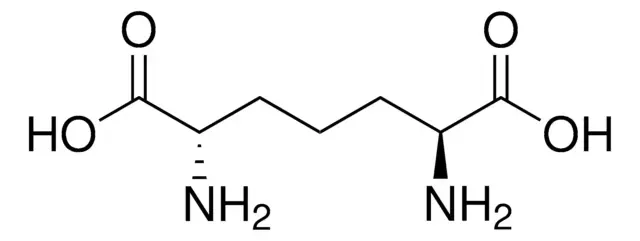
2,6-Diaminopimelic acid is defined by its symmetrical structure: a seven-carbon dicarboxylic acid backbone with amino groups at both the C2 and C6 positions. The IUPAC name, 2,6-diaminoheptanedioic acid, accurately describes this arrangement.

The presence of two stereogenic centers gives rise to three possible stereoisomers: the LL-isomer (both chiral centers in the L-configuration), the DD-isomer (both in the D-configuration), and the meso-isomer (one center L, one center D). This stereochemical diversity is biologically critical, as different isomers serve distinct functions in bacterial metabolism and cell wall architecture.

Natural Occurrence and Biosynthetic Context

DAP is a naturally occurring amino acid found in both bacteria and higher plants. Its discovery by Work in 1950 marked an important milestone in understanding bacterial cell wall composition. In nature, DAP is biosynthesized through the diaminopimelate pathway, which converts pyruvate and L-aspartate into L-lysine, with meso-DAP serving as the immediate precursor to lysine.

The pathway involves several enzymes that have become targets for antibiotic development, including diaminopimelate epimerase (DapF), which catalyzes the interconversion of LL-DAP and meso-DAP, and diaminopimelate decarboxylase, which converts meso-DAP to L-lysine.

Find out more about peptide synthesis here.

Biological Significance and Therapeutic Relevance of DAP

Essential Role in Bacterial Peptidoglycan

The most critical biological function of DAP lies in its contribution to bacterial cell wall integrity. In Gram-negative bacteria, the meso-DAP isomer is incorporated into the peptidoglycan layer, where it acts as a cross-linking agent between glycan strands. Specifically, meso-DAP in the pentapeptide of peptidoglycan provides the attachment site that links the inner and outer membranes to the rigid cell wall structure.

This function is absolutely essential for bacterial survival; disruption of DAP incorporation or biosynthesis leads to weakened cell walls and osmotic lysis. Importantly, because mammals lack DAP and its metabolic pathway, inhibitors targeting DAP utilization exhibit minimal mammalian toxicity, fulfilling the ideal criteria for antibiotic development.

The DAP/Lysine Biosynthetic Pathway as an Antibiotic Target

The diaminopimelate pathway represents a validated target for novel antibacterial agents . Multiple enzymes in this pathway have been structurally characterized and exploited for inhibitor design:

Enzyme TargetFunctionInhibitor ExamplesInhibition Effect
Diaminopimelate Epimerase (DapF)Converts LL-DAP to meso-DAPAziridino-DAP analoguesExtremely potent inhibition; disrupts peptidoglycan synthesis 
meso-DAP Dehydrogenase (m-Ddh)Converts THDP directly to meso-DAPIsoxazoline-containing DAP derivativesSignificant inhibitory potency 
DAP DecarboxylaseConverts meso-DAP to L-lysineα-(Halomethyl)diaminopimelic acidsCompetitive inhibition of lysine biosynthesis 
DAP Aminotransferase (DAP-AT)Transamination step in pathwayHydrazino-dipeptide analoguesSlow-binding inhibition; antimicrobial activity 
MurE (meso-DAP-adding enzyme)Incorporates meso-DAP into peptidoglycan precursorPhosphinate inhibitorsDisrupts peptidoglycan assembly 

Research has demonstrated that certain DAP analogues achieve up to 75% growth inhibition against bacterial strains, validating this approach for antibiotic discovery.

DAP Synthetic Approaches and Peptide Applications

Stereocontrolled Synthesis of DAP-Containing Peptides

The incorporation of DAP into synthetic peptides presents both opportunities and challenges due to its two amino groups and stereochemical complexity. Researchers have developed sophisticated stereocontrolled synthetic strategies to access DAP-containing peptides with defined configurations.

One elegant approach utilizes a chiral synthon derived from L-valine, specifically, a mono-lactim ether that serves as a template for constructing the DAP framework . Alkylation with dihaloalkanes followed by elaboration yields optically active tripeptides with the general structure Val-(DAP)-Val, representing unusual peptides that are C-terminal at both ends of the chain. These compounds are valuable for studying structure-activity relationships and enzyme inhibition.

Complex Peptide Architectures

Beyond simple linear peptides, DAP enables the construction of conformationally constrained structures with diverse biological activities. Researchers have synthesized peptides incorporating a proline residue fused to a diketopiperazine ring containing the DAP skeleton. These fused ring systems resemble natural products such as brevianamides, which exhibit immunomodulatory, antitumor, and antibiotic properties.

X-ray crystallography and computational modeling of these DAP-containing peptides have revealed important structural features, including the planar conformation of diketopiperazine rings and the influence of proline residues on overall molecular geometry. Such insights guide the rational design of peptide-based therapeutics.

Biologically Active Peptide Analogues

Several classes of DAP-containing peptides have demonstrated promising biological activities:

  • γ-Glutamyl-diaminopimelic acid dipeptides: Synthesized via regioselective ring-opening strategies, these compounds show potential as immunostimulating agents .
  • Hydrazino-dipeptide analogues: Designed as inhibitors of DAP aminotransferase, these compounds exhibit potent, slow-binding inhibition and antimicrobial activity against both Gram-positive and Gram-negative bacteria.
  • Phosphonate-containing DAP peptides: While the phosphonate analogues themselves showed weak enzyme inhibition, certain tripeptide derivatives demonstrated growth inhibition against specific bacterial strains.
  • Acylated DAP peptides: Conjugation of DAP-containing peptides with lauric or palmitic acid yields compounds with immunoadjuvant activity.

Research Applications and Custom Synthesis

Tools for Studying Bacterial Enzymology

DAP and its analogues serve as indispensable tools for probing bacterial enzyme mechanisms. Researchers studying DAP epimeraseDAP dehydrogenase, and MurE rely on synthetic DAP-containing substrates and inhibitors to elucidate catalytic mechanisms and to screen for potential antibiotics. The availability of fluorinated, aziridino, and phosphonate derivatives enables detailed structure-activity relationship studies.

Availability from Commercial Sources

For research applications, high-quality DAP and custom DAP-containing peptides are available from specialized suppliers. LifeTein offers expertise in incorporating unusual amino acids like DAP into custom peptide sequences, enabling researchers to access complex structures for their specific investigations. These services include:

  • Synthesis of DAP-containing peptides with defined stereochemistry
  • Incorporation of DAP into cyclic peptides and constrained structures
  • Production of DAP analogues for enzyme inhibition studies
  • Rigorous quality control, including HPLC and mass spectrometry validation

Find out about high-speed RUSH synthesis.

Frequently Asked Questions (FAQ)

What is the difference between the three stereoisomers of DAP?

2,6-Diaminopimelic acid exists as LL-, DD-, and meso-isomers due to its two chiral centers. The LL-isomer has both amino groups in the L-configuration, the DD-isomer has both in the D-configuration, and the meso-isomer has one L and one D center. The meso-DAP isomer is the biologically relevant form incorporated into bacterial peptidoglycan and serves as the immediate precursor to L-lysine.

Why is DAP considered a target for antibiotic development?

Mammals lack the entire diaminopimelate pathway and require dietary lysine, whereas bacteria depend on this pathway to synthesize both meso-DAP (for peptidoglycan) and L-lysine (for protein synthesis). Consequently, inhibitors targeting DAP biosynthesis enzymes or DAP incorporation into peptidoglycan exhibit selective antibacterial activity with minimal mammalian toxicity.

What types of DAP analogues have shown antibacterial activity?

Several classes of DAP analogues demonstrate promising antibacterial effects, including fluorinated derivativesaziridino-DAP (extremely potent against DAP epimerase), isoxazoline-containing analoguesphosphonate derivatives, and hydrazino-dipeptides. Some analogues achieve up to 75% growth inhibition against bacterial strains.

How is DAP incorporated into synthetic peptides?

DAP incorporation requires orthogonal protecting group strategies due to its two amino groups. Researchers typically use stereocontrolled synthesis starting from chiral synthons like mono-lactim ethers derived from valine. Specialized peptide synthesis providers such as LifeTein offer custom synthesis of DAP-containing peptides with defined stereochemistry and high purity.

Paradisi, F., Porzi, G., & Sandri, S. (2001). A new stereocontrolled synthesis of uncommon tripeptides derived from 2,6-diaminopimelic acid (2,6-DAP). Tetrahedron: Asymmetry, 12(23), 3319–3324. https://doi.org/10.1016/s0957-4166(02)00002-2

Galeazzi, R., Garavelli, M., Grandi, A., Monari, M., Porzi, G., & Sandri, S. (2003). Unusual peptides containing the 2,6-diaminopimelic acid framework: Stereocontrolled synthesis, X-ray analysis, and computational modelling. Part 2. Tetrahedron: Asymmetry, 14(17), 2639–2649. https://doi.org/10.1016/s0957-4166(03)00541-x

Chatterjee, B., Mondal, D., & Bera, S. (2021). Diaminopimelic acid and its analogues: Synthesis and biological perspective. Tetrahedron, 100, 132403. https://doi.org/10.1016/j.tet.2021.132403

Cy7 Fluorescent Labeling

Cy7
Cy7 Maleimide

Fluorescent labelling with Cy7 has emerged as a cornerstone technology in advanced biomedical imaging, enabling researchers to visualize biological processes with unprecedented depth and clarity. As a member of the heptamethine cyanine dye family, Cy7 is characterized by its exceptional near-infrared (NIR) fluorescence properties, with excitation and emission maxima at approximately 749 nm and 776 nm, respectively. This spectral positioning within the NIR optical window (650–900 nm) minimizes interference from endogenous biomolecules like hemoglobin and water, allowing for deep tissue penetration of up to 15 cm and significantly reducing background autofluorescence. The strategic application of Cy7 labelling has revolutionized fields ranging from in vivo imaging and photodynamic therapy to drug delivery systems, with recent advances even witnessing a Cy7-based theranostic agent enter clinical trials. Understanding the principles, methodologies, and applications of Cy7 conjugation is therefore essential for researchers seeking to leverage this powerful tool in their investigations.


Key Takeaways

  • Cy7 exhibits excitation/emission maxima at ~749/776 nm, placing it within the ideal NIR window for deep-tissue imaging with minimal background interference.
  • The dye’s heptamethine structure enables dual functionality as both a fluorescent probe and a photosensitizer for photodynamic and photothermal therapies.
  • Conjugation typically employs NHS ester chemistry targeting primary amines or maleimide chemistry for thiol-specific labelling, with reaction conditions carefully optimized to preserve biomolecular activity.
  • Cy7-labelled peptides and proteins are indispensable tools in in vivo imagingtheranostic agent development, and studies of cell-cell interactions such as the LIPSTIC technique.

Fundamentals of Cy7 Structure and Photophysics

Chemical Architecture of Heptamethine Cyanine Dyes

The molecular structure of Cy7 is defined by a central conjugated polymethine chain connecting two nitrogen-containing indole heterocycles. This heptamethine framework creates an extensive π-conjugated system responsible for the dye’s strong absorption in the NIR region. One indole moiety carries a positive charge, resulting in a delocalized cationic structure that influences both the dye’s photophysical behavior and its interaction with biological environments. This unique architecture not only confers exceptional brightness but also enables structural modifiability at multiple sites, allowing researchers to fine-tune properties such as water solubility, targeting specificity, and photosensitizing efficiency.

Spectral Advantages and the NIR Optical Window

The placement of Cy7’s fluorescence within the NIR region is of paramount biological significance. Between 650 nm and 900 nm, light absorption by hemoglobin, water, and lipids is minimal, creating a “therapeutic window” where photons can penetrate tissues deeply without significant attenuation. Consequently, Cy7-labelled probes can be visualized through several centimeters of tissue, making them ideal for whole-animal imaging studiesintraoperative guidance, and deep-tumor visualization. Furthermore, the absence of endogenous NIR fluorescence in most biological specimens ensures exceptionally low background signals, dramatically improving the signal-to-noise ratio in imaging experiments.

Find out more about fluorescent peptides here.

Conjugation Chemistry and Methodologies

NHS Ester Chemistry for Amine Labelling

The most prevalent strategy for Cy7 conjugation targets primary amine groups present on lysine residues or protein N-termini. This approach utilizes Cy7-NHS ester derivatives, where the N-hydroxysuccinimide moiety acts as a leaving group upon nucleophilic attack by the amine. The reaction proceeds efficiently under mild, weakly alkaline conditions (pH 7.4–8.5), forming a stable amide bond that covalently links the dye to the biomolecule. Researchers must carefully control the molar ratio of dye to protein, typically ranging from 3:1 to 10:1, to achieve optimal labelling density while avoiding excessive modification that could compromise biological function.

Maleimide Chemistry for Thiol-Specific Conjugation

For applications requiring site-specific labelling, maleimide-functionalized Cy7 offers an elegant solution by targeting the thiol groups of cysteine residues. This Michael addition reaction proceeds rapidly under physiological conditions and provides positional control when cysteines are strategically introduced into peptide sequences. In some cases, mild reduction may be necessary to expose previously oxidized or disulfide-bonded thiols before conjugation.

Reaction Optimization and Purification

Successful Cy7 labelling demands meticulous attention to reaction parameters. Temperature control, typically maintained at 4–25°C, prevents protein denaturation while ensuring adequate reaction kinetics. Light protection throughout the procedure is essential to prevent photodegradation of the dye. Following conjugation, removal of unreacted dye is accomplished through gel filtration chromatographydialysis, or HPLC purification, yielding high-purity conjugates suitable for sensitive biological applications.

Cy7

Applications in Biomedical Research

In Vivo Imaging and Biodistribution Studies

Cy7’s deep-tissue imaging capabilities have made it indispensable for tracking biodistributiontumor targeting, and pharmacokinetics in living animals. Fluorescently labelled peptides and proteins administered to murine models can be non-invasively monitored over time, providing real-time insights into accumulation patterns at target sites. For example, Cy7-conjugated LPETGG peptides have been employed to visualize immune cell interactions in preclinical cancer models, leveraging the dye’s NIR emission to penetrate through tissues and reveal dynamic cellular processes.

Photodynamic and Photothermal Therapy

Beyond imaging, certain Cy7 derivatives function as potent photosensitizers for cancer therapy. Upon NIR light activation, these molecules generate reactive oxygen species (ROS) or heat, inducing apoptosis in targeted tumor cells. Recent innovations have produced asymmetric Cy7 dyes with remarkably high singlet oxygen quantum yields (ΦΔ up to 1.84), enabling effective photodynamic therapy at previously unattainable depths. Importantly, these agents exhibit cancer cell specificity by leveraging microenvironmental features such as elevated viscosity, while demonstrating negligible dark cytotoxicity.

Studying Cell-Cell Interactions with LIPSTIC

In immunology research, Cy7-labelled LPETGG peptides have proven instrumental in the LIPSTIC (Labelling Immune Partnerships by SorTagging Intercellular Contacts) technique. This elegant method uses bacterial sortase A to enzymatically transfer fluorescent dyes from the LPETGG substrate onto interacting cell surfaces, enabling researchers to track dynamic immune partnerships in vivo and in vitro with single-cell resolution. Such applications underscore the versatility of Cy7 beyond simple structural labelling.

Selecting Cy7 for Your Research

Advantages Over Shorter-Wavelength Dyes

When compared to visible-light fluorophores like Cy3 or fluorescein, Cy7 offers distinct advantages for whole-animal studiesdeep-tissue imaging, and multiplexing experiments where spectral separation is required. Its NIR emission avoids overlap with common fluorescent proteins and organic dyes, facilitating multicolor panels.

Availability from Commercial Sources

High-quality Cy7 derivatives and pre-labelled peptides are readily available from specialized suppliers. LifeTein, for example, offers Cy7 conjugation on custom peptides such as the LPETGG motif, providing researchers with versatile tools for sortase-mediated labelling and imaging applications. These products undergo rigorous quality control, including HPLC and mass spectrometry validation, ensuring reproducibility in demanding experiments.

Find out more about peptide synthesis here.

Frequently Asked Questions (FAQ)

What are the exact excitation and emission maxima for Cy7?

Cy7 exhibits peak excitation at approximately 749 nm and peak emission at approximately 776 nm, placing it squarely within the near-infrared window optimal for deep-tissue imaging.

How does Cy7 compare to Cy5 or Cy5.5 for in vivo work?

While Cy5 (670 nm emission) and Cy5.5 (701 nm emission) are excellent for many applications, Cy7’s longer wavelength offers superior tissue penetration and lower background due to reduced scattering and absorption by endogenous chromophores. The choice depends on the required depth of imaging and compatibility with available instrumentation.

What conjugation chemistries are available for Cy7?

Cy7 is commonly supplied as an NHS ester for amine coupling or as a maleimide derivative for thiol-specific labelling. The NHS ester is preferred for lysine residues and N-termini, while maleimide enables site-specific conjugation to engineered cysteine residues. Site-specific conjugation via click chemistry is also an option using methods such as Cy7-DBCO and a lys(N3) residue.

Can Cy7 be used for photodynamic therapy?

Yes, certain Cy7 derivatives function as effective photosensitizers, generating reactive oxygen species upon NIR light activation. Recent advances have produced dyes with exceptionally high singlet oxygen quantum yields, making them suitable for photodynamic ablation of tumors.

How stable are Cy7-labelled peptides during storage?

Cy7 conjugates should be protected from light and stored desiccated at -20°C for long-term stability. Reconstituted materials may be stored for up to two weeks at -20°C in aliquots to avoid repeated freeze-thaw cycles.

Are Cy7-labelled peptides available commercially for research?

Yes, specialized providers such as LifeTein offer custom synthesis of Cy7-labelled peptides with high purity (>95%) and rigorous analytical validation. These products are suitable for in vivo imaging, flow cytometry, and advanced techniques like LIPSTIC.

Long, L., Cao, X., Shi, X., Zhang, J., & Shi, C. (2025). Modifications and applications of heptamethine cyanine (Cy7) dyes as near-infrared photosensitizers. Coordination Chemistry Reviews, 541, 216780. https://doi.org/10.1016/j.ccr.2025.216780

Khaikate, O., Muangsopa, P., Piyanuch, P., Khrootkaew, T., Wiriya, N., Chansaenpak, K., Sukwattanasinitt, M., & Kamkaew, A. (2024). Asymmetric heptamethine cyanine dye for viscosity detection and photodynamic therapy. Journal of Photochemistry and Photobiology A: Chemistry, 453, 115659. https://doi.org/10.1016/j.jphotochem.2024.115659