Acetylation was for a long time the only acyl mark anyone looked for on a lysine. It is no longer alone. Alongside acetylation, eight further short-chain lysine acylations have been established as histone modifications — propionylation, butyrylation, 2-hydroxyisobutyrylation, succinylation, malonylation, glutarylation, crotonylation and β-hydroxybutyrylation — each regulated by enzymatic and metabolic mechanisms and each with physiological functions of its own.1 Lactylation was added to the list in 2019.2
What makes the series interesting is where the acyl groups come from. The donors are acyl-CoA species drawn from central metabolism — crotonyl-CoA, succinyl-CoA, lactoyl-CoA — so the acylation state of chromatin reflects the metabolic state of the cell, and the modifications act as a route by which metabolism reaches gene expression.1
We make peptides carrying any of these marks at a defined position. Below is what each one weighs, where the analytical traps are, and what we need from you to build one.
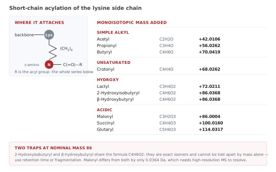
Most work on these marks is done with antibodies, and antibody quality in this field has a documented history of trouble. A survey of more than 200 antibodies raised against 57 histone modifications found that over a quarter failed specificity testing by dot blot or western blot, and that among those that did pass, more than a fifth failed in chromatin immunoprecipitation.3 A later peptide-microarray study of more than 100 commercial histone antibodies made the same point in more detail and put the results in a public database.4
The reason is structural rather than careless manufacturing. The chemical differences between these marks are small, and the sequence around them is often repetitive — H3K9 and H3K27 both sit in an identical ARKS motif, so an antibody has to read the residues outside that motif to tell the two sites apart.5 Add the near-isobaric pairs in the figure above and it becomes clear why an antibody raised against one acylation may also report a different one.
A synthetic peptide carrying one mark at one known position is the control that settles it. Peptide arrays are the established format for profiling histone antibody specificity,4 and the same peptides serve as blocking competitors, as binding partners for reader-domain pull-downs, and as standards that fix retention time and fragmentation behaviour in a mass spectrometry method.
| Class | Modifications | Typical use |
|---|---|---|
| Simple alkyl | Acetyl, propionyl, butyryl, isobutyryl, hexanoyl | Reader-domain binding, HDAC and sirtuin substrate work |
| Unsaturated | Crotonyl | YEATS and bromodomain binding, antibody validation |
| Hydroxy | 2-Hydroxyisobutyryl, R- and S-3-hydroxybutyryl, lactyl | Metabolic chromatin studies, MS standards |
| Acidic | Malonyl, succinyl, glutaryl | Charge-reversal studies, sirtuin assays |
| Aromatic and other | Nicotinyl, isonicotinyl, benzoyl, dopaminyl, serotonyl | Less common marks, usually as antibody controls |
| Methylation | Mono-, di- and tri-methyl lysine; symmetric and asymmetric dimethyl arginine | Frequently ordered alongside acylations on the same sequence |
All of these can be combined with the formats that make a peptide usable in an assay: an N-terminal or C-terminal biotin for array printing and pull-down, a spacer between the biotin and the sequence, a fluorescent label, or a free versus capped terminus. See the full catalog entry for the complete list of building blocks.
Four things decide whether the peptide you receive is the one your experiment needs:
H3K9cr rather than “crotonylated H3”.Where a project calls for a set — one mark per peptide across a panel, or the same mark walked along a sequence — we build them together so that they share purification and analysis conditions.
The mass shifts in the figure are monoisotopic and additive, so a peptide carrying two marks shows the sum. Two cases need care, and both are visible in the table above: 2-hydroxyisobutyryl and β-hydroxybutyryl are exact isomers, identical in formula and mass, separable only by retention time or fragmentation; and malonyl sits 0.0364 Da away from both, which is within reach of a high-resolution instrument but not of a modest one.
The same 0.0364 Da gap separates trimethylation from acetylation, which is the better-known version of the same trap. Our modification mass table covers that pair and the other common near-coincidences.
Tell us the mark and the position. Send the sequence with the modification written in, and say what the peptide is for — an array, a pull-down, an MS standard. If a different format would serve the experiment better, we will say so before quoting. We send a quote within 24 hours.
All LifeTein products are supplied for laboratory research use only. They are not for human or veterinary use, and not for diagnostic or therapeutic use.