


QSY 9 is not a fluorophore. It absorbs strongly near 560 nm and releases that energy as heat rather than light, so its role is to switch off a nearby dye until something separates the two. That is the basis of a fluorogenic peptide substrate.
| Absorbance maximum | 560 nm |
| Emission maximum | None — dark quencher |
| Laser line | — |
| Dye class | Dark quencher |
| Absorbance maximum | About 560 nm |
| Emission | None — a dark quencher |
| Reactive form | NHS ester; maleimide for cysteine |
| Pairs with | Green and orange donors such as FAM, FITC and TAMRA |
| Typical use | Protease substrates and other cleavage reporters |
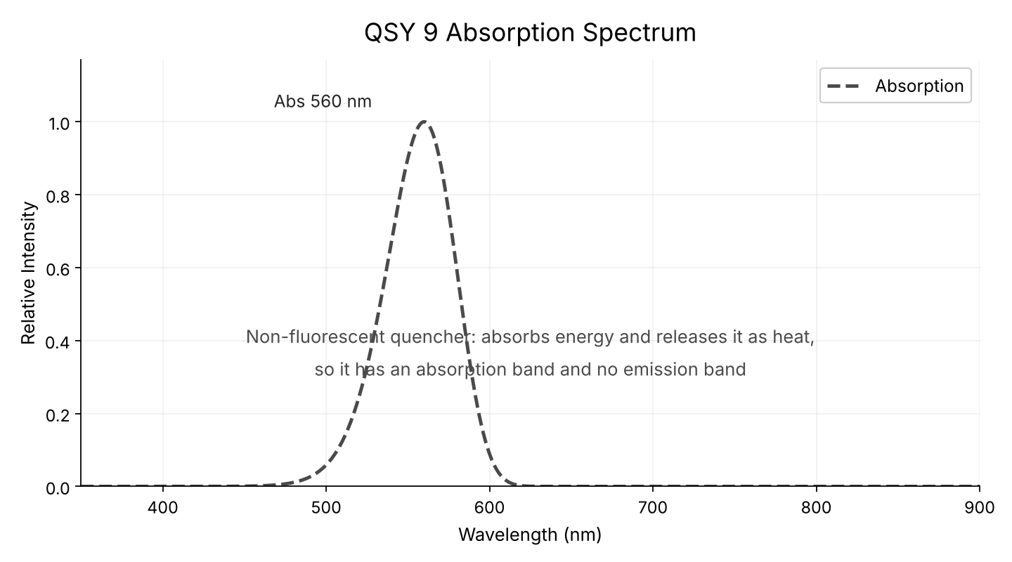
In a quenched substrate the signal of interest is the appearance of fluorescence on cleavage. A fluorescent acceptor would emit its own light, adding a background that has to be separated from the donor. A dark quencher emits nothing, so the intact substrate is genuinely dark and the signal-to-background after cleavage is as large as the chemistry allows.
Quenching efficiency depends on the overlap between the donor's emission and the quencher's absorption, and on the distance between them. QSY 9 absorbs near 560 nm, which overlaps the emission of green donors such as FAM and of orange donors such as TAMRA. For a far-red donor a longer-wavelength quencher is needed instead.
The distance between dye and quencher matters as much as the spectral overlap, so the two are normally placed at opposite ends of the cleavage site with the recognition sequence between them. A substrate that shows high background before cleavage is usually too long between the pair, or is aggregating; both are worth checking before concluding the enzyme is inactive.
| Situation | Choice | Why |
|---|---|---|
| Protease substrate with a FAM or TAMRA donor | QSY 9 | Absorption overlaps both donors |
| Far-red donor | A longer-wavelength quencher | Overlap must match the donor emission |
| Reporter that must emit in both states | A second fluorophore instead | A dark quencher never emits |
| Ratiometric FRET measurement | A fluorescent acceptor | Ratiometric reads need two emission signals |
Tell us what the peptide has to do in the assay as well as its sequence. Where the dye sits often matters more to the result than which dye it is, and a label placed in a binding motif can remove the activity being measured.