Curli and HSV-1 Antibody Correlation Shown in Parkinson’s Disease Patients

peptides for Parkinson’s Disease

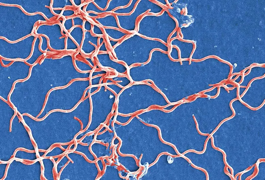
Parkinson’s Disease is an ever-prevalent neurodegenerative disease whose cases are expected to double within the next decade. While it is a multifactorial disorder that may stem from a combination of environmental and genetic factors, a more recent study has shifted the spotlight onto gut microbiota and microbial pathogens as a large contributor to neurodegenerative diseases altogether. Specifically, curli-producing bacteria in the gut microbiota could increase and facilitate alpha-synuclein aggregation in the brain. This spearheaded some research into the Curli and HSV-1 antibody correlation shown in Parkinson’s Disease patients.

Curli-producing bacteria in the gut could lead to neurodegenerative diseases

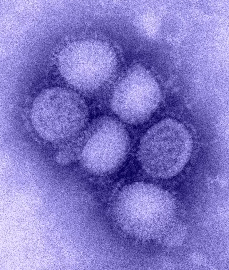
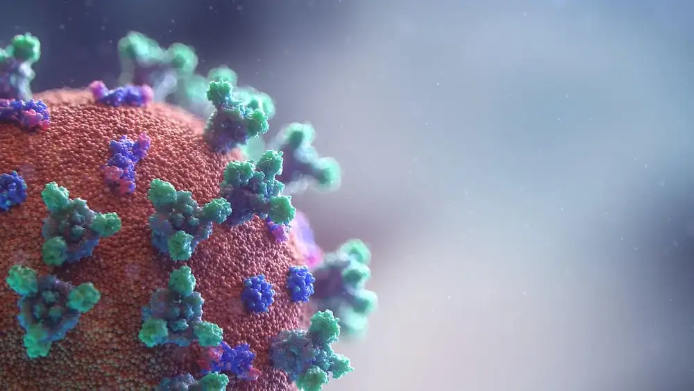
LifeTein supplied the group pursuing this work with an immunogenic peptide derived from bacterial amyloid curli to help test their theory. They chose to analyze the humoral response against this peptide as well as other α-syn peptides in both Parkinson’s Disease patients and healthy controls. What was found was a significant correlation in the Parkinson’s Disease patients’ humoral response against curli and HSV-1 when compared to the control.

While the study shows this high correlation exists between these bacterial peptides and Parkinson’s Disease, it is only the first step in this journey. The role of each of these pathogens in Parkinson’s Disease is yet to be explored and still offers a new angle of understanding the neurodegenerative disease, and hopefully a path for treating it as well.

Jasemi S, Paulus K, Noli M, Simula ER, Ruberto S, Sechi LA. Antibodies against HSV-1 and Curli Show the Highest Correlation in Parkinson’s Disease Patients in Comparison to Healthy Controls. International Journal of Molecular Sciences. 2022; 23(23):14816. https://doi.org/10.3390/ijms232314816

The Vital Role of 14-3-3γ in Influenza A Virus Replication

Influenza A is a virus responsible for multiple pandemics over the last centuries, the respiratory disease has claimed millions of lives over the course of human history. Though other pandemics may come to mind in recent years, flu season is just starting up again now as the weather gets chillier. While we can fight back the virus on a yearly basis, more understanding is requited for a long-term victory. A team has been researching into the NS1 protein of the virus, the part responsible for downregulating the antiviral response of host cells to facilitate viral replication. They believe their work has revealed information on the vital role of 14-3-3γ in influenza A virus replication, where the isoform was found to interact with the protein.

Truncated N-Terminus interacts with 14-3-3γ

The group preformed much work such through immunoprecipitation to show the interactions between 14-3-3γ and the influenza A encoded NS1 protein. Some of their most compelling finds was the inhibition of 14-3-3γ expression in the host cells greatly reduced replication of the PR8 wild-type virus, but had no such effect on the R8-NS1/1-98 mutant virus, which lacks most of the effector domain of NS1. LifeTein was able to provide the group with anti β-tubulin antibodies, which assisted in their immunoprecipitation methods.

The team insists that the evidence points directly towards the vital role of 14-3-3γ in influenza A virus replication thanks to the NS1 protein. While they are still unclear on the precise mechanisms of these interactions, they are certain the findings have laid out the groundwork for future pivotal studies involving influenza A and the role of 14-3-3γ in infection.

Kuo, R.-L.; Tam, E.-H.; Woung, C.-H.; Hung, C.-M.; Liu, H.-P.; Liu, H.M.; Wu, C.-C. Interactome Profiling of N-Terminus-Truncated NS1 Protein of Influenza A Virus Reveals Role of 14-3-3γ in Virus Replication. Pathogens 2022, 11, 733. https://doi.org/10.3390/pathogens11070733

Peptides Fold and Self-Assemble on Graphite-Water Interfaces

J. Chem. Inf. Model. 2022, 62, 17, 4066-4082

The concept of self-assembling peptides is a promising front where construction of devices can be achieved through a single molecule. While the outcome is enticing, the means to reach a consistent outcome are complex to say the least. Dozens of factors go into how a peptide may self-assemble and fold, with the most important being the sequence itself. While this can be handled by careful screening and simulations, the interface at which this folding occurs becomes more important to consider at well. Researchers looked to test how specific peptides fold and self-assemble on graphite-water interfaces, where a number of factors give this method the advantage over doing so in free solution.

Graphite helps peptides self fold into conformations

The group studying this phenomenon claimed that the folded conformations of the peptides were stable over a variety of temperatures when observed over graphite. They point out that it is due to the peptide backbone aligning with the zigzag directions of the graphite plane, thus allowing the conformations to occur more favorably from the intermolecular hydrogen bonds of the molecule. Atomic force microscopy revealed these theories to be true beyond initial simulations as well.

The team believes the design principles displayed in these experiments could be of great use in future iterations of self-assembling peptide engineering. The thermodynamically favored self-assembly with the use of a graphite-water interface shows promise as a medium for even more complex molecular devices in the future, a future LifeTein is looking forward to being a part of.

Justin Legleiter, Ravindra Thakkar, Astrid Velásquez-Silva, Ingrid Miranda-Carvajal, Susan Whitaker, John Tomich, and Jeffrey Comer
Journal of Chemical Information and Modeling 2022 62 (17), 4066-4082
DOI: 10.1021/acs.jcim.2c00419

The Nobel Prize in Chemistry 2022 awarded to Carolyn R. Bertozzi, Morten Meldal, and K. Barry Sharpless for their outstanding work with Click Chemistry

The Nobel Prize in Chemistry 2022 awarded to Carolyn R. Bertozzi, Morten Meldal, and K. Barry Sharpless for their outstanding work with Click Chemistry
Peptide Click Chemistry

The Nobel Prize in Chemistry 2022 awarded to Carolyn R. Bertozzi, Morten Meldal, and K. Barry Sharpless for their outstanding work with click chemistry, a well-deserved honor for developing a vital and straightforward technique in modern chemistry. Barry Sharpless, with this as his second Nobel Prize in Chemistry, and Morten Meldal independently presented the idea of click chemistry over twenty years ago. The reactions involved are highly versatile and can be performed under a multitude of conditions, making the method incredibly efficient and applicable to many fields. In a perfect example, Carolyn Bertozzi, who was jointly awarded this Nobel Prize, utilized the principles of click chemistry to present bioorthogonal chemistry, click reactions that take place within living organisms without disrupting the normal chemistry of the cell. Since its inception, click chemistry has had its influence explored across many other facets of chemistry, including very useful applications in peptide synthesis.

LifeTein frequently uses this CuAAC reaction in its oligonucleotide-peptide conjugation service. Such reactions would not be nearly as feasible without the simple answer of click chemistry, as linking large and functionalized molecules like peptides and oligonucleotides is a very challenging task otherwise. With this, LifeTein is able to provide DNA-peptide conjugates and RNA-peptide conjugates for cell screening and in vivo studies with ease.

Congratulations again to Carolyn R. Bertozzi, Morten Meldal, and K. Barry Sharpless on their joint award for The Nobel Prize in Chemistry 2022, the field wouldn’t be anywhere near where it is today without click chemistry.

Our Click Chemistry Products and Services:

Custom Oligo-peptide Conjugate Service Using Click Chemistry

Custom Pegylation Service Using Click Chemistry

Linear Peptide Epitomes to Better Diagnose Lyme Disease

Lyme disease is an abundantly present vector-born disease, with nearly 400,000 new cases in the U.S. diagnosed each year. While treatment with antibiotics is effective, if the disease goes unnoticed and unchecked it can lead to lifelong damage to the nervous and musculoskeletal system. Hence, it is critical to detect the disease as early as possible, however this becomes a complex issue since not every case results in erythema migrans (EM), the characteristic skin lesion of Lyme disease. This issue is further complicated since current serodiagnostics lack sensitivity in early Lyme disease detection, when treatment would be the most effective. One group sought to solve this issue by developing a method more sensitive and more specific than the current ones, ultimately using linear peptide epitomes to better diagnose Lyme disease.

Linear peptide epitomes lead to higher specificity and sensitivity in Lyme disease assays

LifeTein synthesized peptides from prominent B. burgdorferi antigens expressed during human infection of Lyme disease for the group to utilize. Their idea is to use synthetic peptides that contain the linear epitomes necessary, rather than protein antigens that often have cross-reactive epitomes. Eliminating the cross-reactive epitomes leads to more specificity within the tests, and more sensitivity as well. While the results for the single peptide epitomes were less than anticipated, the group found that using the epitome peptides in pairs resulted in the specificity and sensitivity they had hypothesized.

Diagnostic assessment of Lyme disease is in dire need of an improvement to efficiently catch it in the early stages, and these linear peptide epitomes are shaping to be a key part of the solution. The group hopes that their research will allow future works to improve upon their studies and one day include the linear peptide epitomes as part of a potential multi-peptide-based assay for the laboratory diagnosis of Lyme disease.

Arnaboldi PM, Katseff AS, Sambir M, Dattwyler RJ. Linear Peptide Epitopes Derived from ErpP, p35, and FlaB in the Serodiagnosis of Lyme Disease. Pathogens. 2022; 11(8):944. https://doi.org/10.3390/pathogens11080944

Higher HERV-W and IFN-I Antibody Presence Detected in Intensive Care COVID-19 Patients

Higher HERV-W and IFN-I Antibody Presence Detected in Intensive Care COVID-19 Patients

As the world moves on through the ongoing pandemic that is COVID-19, great minds across every field and corner of the world are doing their part to further our collective understanding of the virus. A recent study specifically took greater notice in the population of antibodies increasing in more sever cases of COVID-19 over mild ones. Specifically, there was a study conducted on the higher HERV-W and IFN-I antibody presence detected in intensive care COVID-19 patients, utilizing peptides for the experiment.

HERV-W and IFN-I antibody presence higher in severe COVID-19 cases

LifeTein supplied the group with the HERV-W-env(248–262), IFN-α, and IFN-ω peptides necessary for the report. Not only did the studies prove higher levels of anti-IFN-I autoantibodies and HERV-W antibodies are much more prevalent in severe COVID-19 cases than in mild ones, but it opened up a new perspective on how the humoral responses against HERV-W-env(248–262) and IFN-α are correlated across ICU patients. Hopefully, this research leads to more beneficial breakthroughs over time, for COVID-19 cases and potentially other life-threatening conditions as well.

Simula, E. R., Manca, M. A., Noli, M., Jasemi, S., Ruberto, S., Uzzau, S., Rubino, S., Manca, P., & Sechi, L. A. (2022). Increased Presence of Antibodies against Type I Interferons and Human Endogenous Retrovirus W in Intensive Care Unit COVID-19 Patients. In H. H. Mostafa (Ed.), Microbiology Spectrum. American Society for Microbiology. https://doi.org/10.1128/spectrum.01280-22

Revolutionary LIPSTIC Method Illuminates Receptor-Ligand Interactions In Vivo And In Vitro

-LIPTSTIC mechanism, from the cited paper.

Cell interaction analysis is a cornerstone of biological research, providing critical insights into the intricate world of molecular communication within living organisms. While traditional microscopy offers a glimpse into these interactions, it often falls short when it comes to revealing the specific receptors and ligands involved. Enter a groundbreaking method known as Labeling Immune Partnerships by SorTagging Intercellular Contacts, or LIPSTIC for short, which has been developed by a team of innovative scientists.

At the heart of LIPSTIC lies the ingenious combination of a fluorescent LPXTG peptide motif and Staphylococcus aureus transpeptidase Sortase A (SrtA), offering a highly effective means of tracking and studying cell interactions. This novel approach is readily detectable through flow cytometry, making it a game-changer in the field of biological research.

The LIPSTIC method hinges on the LPETG peptide and SrtA reaction, a technique that allows for the labeling of receptor and ligand interactions. LifeTein, a leading supplier in the life sciences industry, played a pivotal role by providing the necessary Biotin-ahx-LPETG peptide to the research group. In the LIPSTIC method, a noteworthy ligand or receptor is fused with a tag composed of five N-terminal glycine residues (G5). The SrtA enzyme then graciously donates the fluorescent peptide to this fusion, enabling precise monitoring of the acceptor cell post-separation.

One of the most impressive aspects of LIPSTIC is its versatility. It empowers scientists to analyze cell-cell interactions both in vitro and in vivo, offering a comprehensive understanding of molecular partnerships in various biological contexts. Moreover, LIPSTIC’s sensitivity is a standout feature, as it can even detect rare or low-intensity interactions that might have otherwise remained hidden.

In conclusion, the introduction of the LIPSTIC method marks a significant advancement in the field of cell interaction analysis. Its ability to unveil the intricacies of receptor-ligand interactions in living systems, along with its applicability in diverse research settings, positions LIPSTIC as a powerful tool for scientists striving to unlock the secrets of cellular communication.


Pasqual G, Chudnovskiy A, Tas JMJ, Agudelo M, Schweitzer LD, Cui A, Hacohen N, Victora GD. Monitoring T cell-dendritic cell interactions in vivo by intercellular enzymatic labeling. Nature. 2018 Jan 25;553(7689):496-500. doi: 10.1038/nature25442. Epub 2018 Jan 17. PMID: 29342141; PMCID: PMC5853129.

Mechanism of Cisplatin binding with Oligonucleotides and Peptides

Platinum based anticancer drugs generally go hand-in-hand with DNA, however their interactions with proteins are important to understand as well. A group focused their work on discovering and further exploring binding sites of cisplatin onto both an oligonucleotide and two peptides corresponding to segments of H2A and H2B histone proteins via mass spectrometry. The study revealed key interactions and the mechanism of cisplatin binding with oligonucleotides and peptides, and eventually DNA as well.

Peptide and oligonucleotide competitive binding with platinum

LifeTein supplied the group with two peptides for the project; P1, from the acidic region of H2a, and P2, from the α2-helix of H2B human histone protein. These peptides and an oligonucleotide model of DNA were analyzed using electrospray ionization high-resolution mass spectrometry and tested with substantial theoretical figures to match the fragments. Here the group found the preferred binding sites on both of the peptides and the oligonucleotide, of relevance are the multiple histidine and methionine residues on P2 that saw the most binding to cisplatin over P1.

The adducts formed on platinated P2 were tested and incubated solely with the oligonucleotide, where after two days the group found that a portion of the platinum fragments had migrated from the peptide to the oligonucleotide. This observation not only showed that the oligonucleotide was perhaps the more thermodynamically favored product, but also gave insight into how the cisplatin drug takes to DNA during chemotherapy, by binding to the proteins first and then reversing the reaction and migrating. The long-term implications of this mechanism will surely be researched and explored in the future of platinum drug delivery and cancer treatment.

Mansouri, F., Patiny, L., Ortiz, D. et al. Simultaneous mass spectrometry analysis of cisplatin with oligonucleotide-peptide mixtures: implications for the mechanism of action. J Biol Inorg Chem 27, 239–248 (2022). https://doi.org/10.1007/s00775-022-01924-9

αCT1 Peptide Weakens Cancerous Glioma Cells

Glioblastoma (GBM) is the most commonly occurring terminal brain cancer. Due to complications in the brain like the blood brain barrier, methods of treating GBM are few and far between. Therefore, treatment in the region is generally left to specific chemotherapeutics like temozolomide (TMZ), which has the unique capability to bypass the brain blood barrier. However, matters become more complicated as many subpopulations of GBM, namely the glioma stem cell populations, are resistant to TMZ. Researchers are looking into ways to bypass this resilience, namely connexin 43 (Cx43) hemichannels that when inhibited by mimetic peptides allow the glioma stem cell populations to be treated significantly more effectively by TMZ

Cx43 mimetic peptides weaken cancer’s resistance to TMZ

Researchers used LifeTein’s peptide synthesis service to create two mimetic peptides of Cx43, αCT11 and αCT1, to inhibit Cx43 hemichannels and then sensitize the glioma cells and other GBM cell populations to TMZ in a 3D hyaluronic acid and collagen hydrogel-based tumor organoid system. After testing this model extensively, the group found that only the αCT1 peptide in combination with TMZ proved effective in treating the cell lines. It is believed that the αCT1 is more successful due to its cell penetrating sequence when compared to αCT11.

Overall, the group emphasizes that the model used does not accurately mimic the cellular heterogeneity of GBM, but the results are a fantastic start and can be used as a tool to further study treatment of this aggressive brain cancer. Further work can optimize this treatment and can hopefully provide a chance for those who have to go against this fatal ailment.

Jingru Che, Thomas J. DePalma, Hemamylammal Sivakumar, et al. αCT1 Peptide Sensitizes Glioma Cells to Temozolomide in a Glioblastoma Organoid Platform. Authorea. April 29, 2022.

Unlocking the Antibacterial Potential: Human SCPPPQ1 Protein and Its Derivatives

Localization and effect of SCPPPQ1 on P. gingivalis bacteria using TEM imaging. From the cited paper.

After the gut, the mouth is the most important microbiome in the human body, with the inhabitants ranging from bacteria and fungi to viruses and protozoa. If this careful balance of microorganisms is thrown off even by a little, certain pathogens could propagate and cause serious periodontal diseases. One such pathogen is Porphyromonas gingivalis (P. gingivalis), which degrades the specialized basal lamina components that protect supporting teeth tissues. However, one component of this specialized basal lamina, the secretory calcium-binding phosphoprotein proline-glutamine rich 1 (SCPPPQ1) protein, had shown to be not only resistant to P. gingivalis, but also to affect the cell membrane of P. gingivalis itself. A group of researchers then decided to explore the antimicrobial properties of the SCPPPQ1 protein and its peptide derivatives.

SCPPPQ1 protein and peptide derivatives as antibacterial agents

Using the SCPPPQ1 protein itself and derived peptides synthesized by LifeTein, the group sought to test how they fared against P. gingivalis in isolated conditions. After incubating the two together, results showed a rapid and significant decrease of P. gingivalis population. The means of which were narrowed down to aggregation of bacteria and membrane disruption.

The group went further and tested the antibacterial properties against other pathogens, and though there were results, none were as significant as those against P. gingivalis. The results point towards a more honed treatment against P. gingivalis using this knowledge of the SCPPPQ1 protein and its peptide derivatives. Since the protein itself is native to the human mouth, further application to treat periodontal pathogens with its antibacterial properties is not out of the question.

Mary, C., Fouillen, A., Moffatt, P. et al. Effect of human secretory calcium-binding phosphoprotein proline-glutamine rich 1 protein on Porphyromonas gingivalis and identification of its active portions. Sci Rep 11, 23724 (2021). https://doi.org/10.1038/s41598-021-02661-w