Self-Assembling Peptide Hydrogel as a Versatile Drug Delivery Platform

Peptide Hydrogel

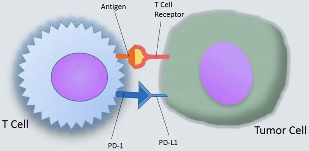
Hydrogels with a capacity to absorb and hold water within a porous, swelled structure make it a great candidate as a drug delivery system due to their broad range of physical properties as well as chemical adaptability. For example, a positively charged polypeptide (poly-l-lysine, PLL) coupling to a self-assembling dipeptide (Fmoc-FF) leads to the formation of hydrogels with rheological properties suitable for injection.

Self-Assembling Peptide Hydrogels As a Drug Delivery System

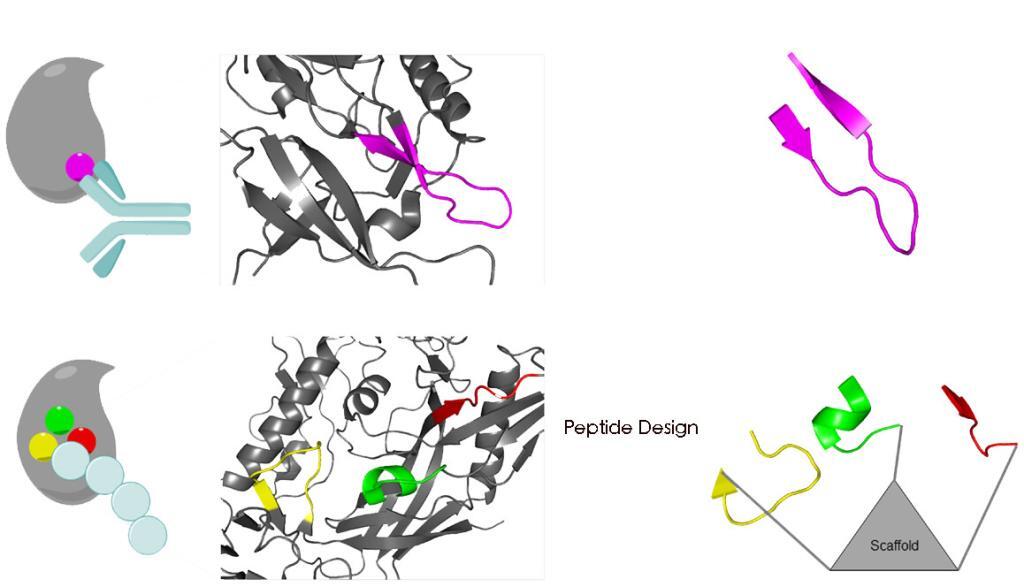
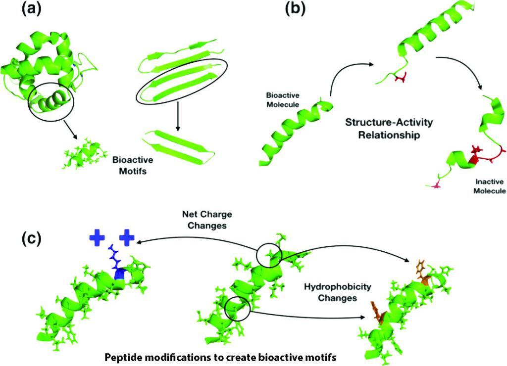
Hydrogenation via self-assembly is a hierarchical process. The hydrogels can be easily prepared via simple mixing and incorporated with various stoichiometric ratios of peptides without any additional synthetic processes. Peptides can form hydrogels by multiple non-covalent interactions. It was found that PTZ-Gly-Phe-Phe-Tyr can form gels at ultra-low concentrations of 0.01 wt.%. The π-π stacking may be another driving forces in the self-assembly to form the final hydrogel structure. The N-cadherin mimic peptide (CLRAHAVDIN) and TGF-β1 mimic peptide (CESPLKRQ) synthesized by LifeTein were used to form an injectable 3D hydrogel. Increased retention of stem cells by using an injectable hydrogel has resulted in successful tissue engineering outcomes.

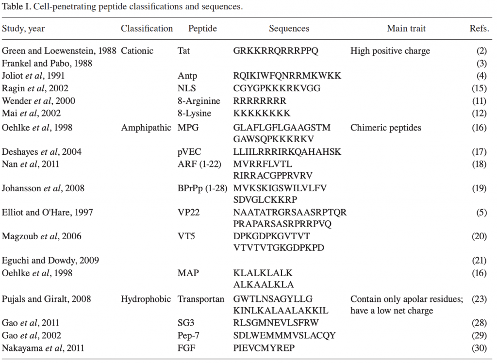
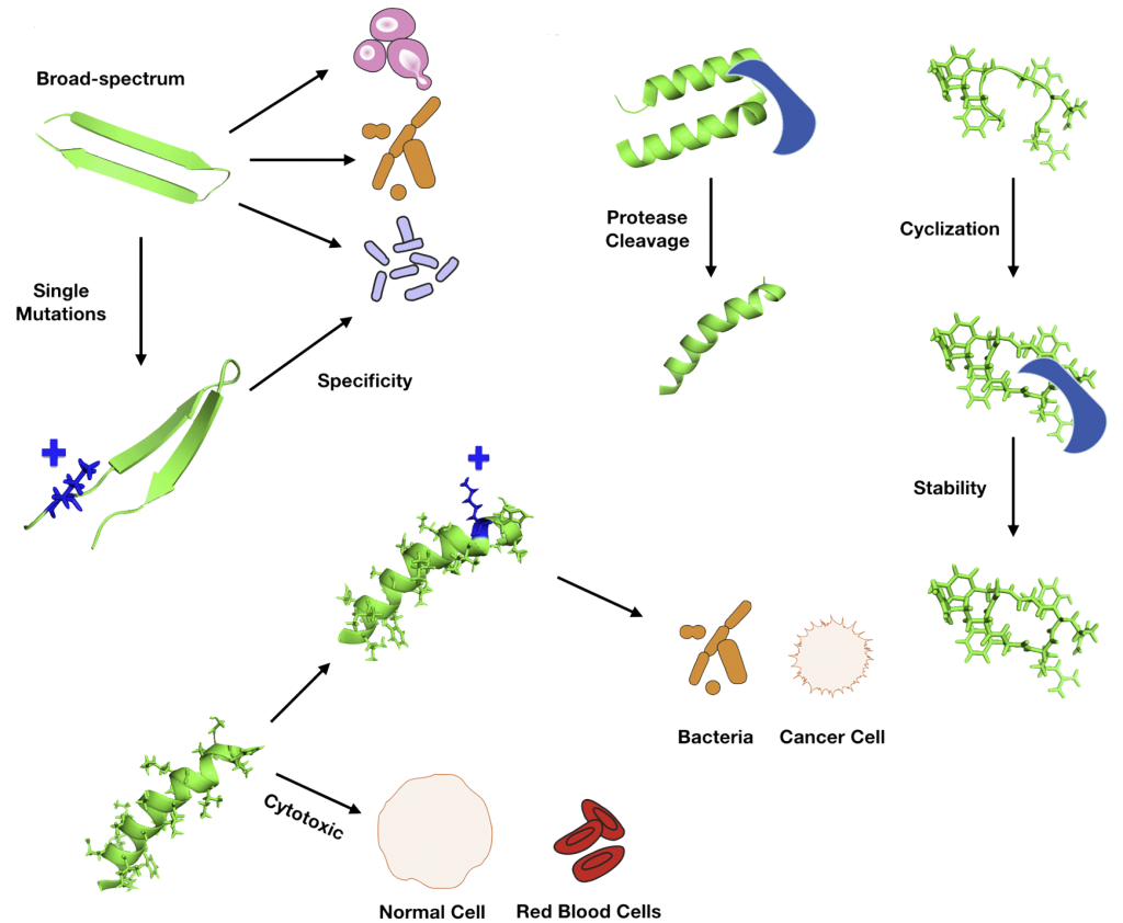
Another efficient method to facilitate hydrogel formation is based on the electrostatic attraction of oppositely charged peptides. The order of amino acids is of great importance for hydrogenation. Self-assembling beta-hairpin peptides, with high arginine content, exhibited extremely good performance in killing bacteria. The peptide hydrogels also have been demonstrated to be used as wound healing agents and other therapeutic instruments under different conditions. Curcumin could be encapsulated into hairpin hydrogels as an injectable agent for localized delivery.

Peptide Synthesis Home Page

Our Services:

COVID-19 Services & Products

Custom Antibody Services

Rush Peptide Synthesis

Peptide Nucleic Acids (PNAs)

Custom Peptide Synthesis Services

Gene Synthesis Service

Custom Chemical Synthesis

Other Posts:

LifeTein peptide FLAG(GS)HA

Smaller Ions Stabilize β-sheets

Synthetic Scorpion Toxin Peptides for Chronic Pain

LifeTein’s Braftide & Cancer Therapy

How to Identify a PD-L1 Binding Peptide for Determination of PDL1 Expression in Tumors

PD-L1 Binding Peptides

Blocking the interaction between Programmed Death Ligand 1 (PD-L1) and its receptor, PD-1, is an effective method of treating many types of cancers.

  1. Identifying the PD-L1 binding peptide and mock peptide: Select from a library of peptides for stability and high affinity.
  2. Fluorescent target peptide-Cy5 as Marker for Flow Cytometry.
  3. Detection of PD-L1 in circulating tumor cells using the Cy5 or Cy7 (infrared) labeled peptides.
  4. Biotinylated peptides to detect PD-L1 in sample tissues. The streptavidin-HRP are used.
  5. Fluorescent Cy5-labeled peptides detects PD-L1 in sample tissues.

By utilizing a peptide-based approach, it is possible to detect all levels of PD-L1 with high sensitivity and specificity.

Reference:

https://www.nature.com/articles/s41598-017-10946-2

How to screen small peptide ligands for targeting receptors in cancer cells?

  1. Design a biotin-labeled peptide library: overlapping or Alanine scan
  2. The peptide library is coated on the avidin-coated 96 well plates.
  3. The Peptide library is used to evaluate the binding of each peptide to different cell lines using CyQUANT Cell Proliferation Assays.
  4. Flow cytometry: Determine the cellular uptake of selected FITC-labeled peptides.
  5. Competitive assay: The cells are incubated with FITC-peptide with or without excess unlabeled peptide (100-fold)
  6. Finetune the peptide sequence: structure analysis, cyclization, and D amino acid modification.
  7. Find the promising receptor-specific cancer cell binding peptide that can be conjugated directly to a chemotherapeutic drug or to nanoparticles for targeted drug delivery to enhance the efficacy of chemotherapy for cancer treatment.

Reference:

https://www.nature.com/articles/s41598-019-38574-y

Synthetic Peptides as Protein Mimics in Biological Research

Synthetic Peptides as Protein Mimics

Synthetic peptides have proven an excellent type of molecule for the mimicry of protein sites. The modified peptides increase the proteolytic stability of the molecules, enhancing their utility for biological applications.

Toolbox for Peptide Synthesis: Non-Proteinogenic Amino Acids and Site-Selective Ligation

The long peptides can be synthesized by the ligation method. Amino acid derivatives with modified backbone length and side-chain orientation, such as d-amino acids, N-alkyl glycine monomers, or proteolytically stable amino acid derivatives can be introduced to the peptides.

Protein Secondary Structure Mimics: α-Helix Mimics, β-Sheet Mimics
Peptide chains can be organized into secondary structures, such as α-helices and β-sheets. Peptides that mimic α-helices and β-sheets of proteins are attractive targets for drug development and tools to explore protein binding mechanism.

The α-helical conformation of a peptide can be induced by adding covalent links between amino acid side chains at selected positions. These links can be formed by lactam and disulfide bridges, triazole-based linkages, and hydrocarbon staples.

In β-sheets, β-strands are connected via loops or turns. Methods to mimic turn structures include macrocyclization, dipeptide of d-proline and l-proline, or α-aminoisobutyric acid in combination with either a d-α-amino acid or an achiral α-amino acid. An example of stimuli-responsive peptides is the temperature-dependent formation of hydrogels by β-sheet peptides. The β-hairpin mimic undergoes gelation upon heating at 60°C, and is completely reversible while cooling.

Protein Mimics in Biomedical Research

Peptides mimicking the CHR region of gp41 were developed to inhibit the formation of the six-helical bundle. Peptides that mimic these receptors are useful tools to explore the details of virus infection mechanism, as well as to develop new drugs against HIV-1. Peptides that mimic the extracellular domains of seven transmembrane G protein-coupled receptors (GPCRs), which is composed of the N-terminus (NT) and the three extracellular loops (ECLs) were explored. Peptide Ac-RERF-NH2 has a high propensity to adopt an α-turn structure and could be a promising drug candidate against cancer.

The design of peptides as protein mimics has evolved as a promising strategy for the exploration of protein-protein interactions, as they are biocompatible, biodegradable, and functionally selective.

Explainer: Why peptides are the ‘next big thing’ in medical research?

Cell Penetrating Peptides for Drug Discovery

Biochemists are excited by the possibilities presented by peptides and proteins as pharmaceuticals because they so often mimic exactly the behavior of a natural ligand – the substance that interacts with the receptor on an enzyme or cell to cause a biological process.

Peptides are used for:
1. Self-Assembling Peptide Epitopes as Novel Platform for Anticancer Vaccination: OVA 250−264 and HPV16 E7 43−57.

2.Incorporation of DSPE-PEG and cRGD-modified DSPE-PEG molecules improves the biocompatibility and cellular uptake of the nanoprodrug platform: Click chemistry conjugation of peptides to PEGs

3. Designing Tracers for Imaging from Cyclic Peptides: ATTO, FITC, FAM, Cy3, Cy5, infrared C7

4. Co-delivery of tumor antigen and dual toll-like receptor ligands into dendritic cell: Cell Penetrating Peptides, HIV-TAT proteins, R8.

How to Design Peptide for Antimicrobial Applications

Antimicrobial Peptides

There are many ways to design improved peptides: site-directed mutagenesis, computational design approaches, synthetic libraries, template-assisted methodologies, and mechanism-based strategies.

  1. Site-directed mutagenesis:
    Adding, deleting, or replacing one or more amino acid residues are useful approaches to re-engineer a peptide. The alanine or lysine scanning are examples of valuable strategies, as they allow for coverage of all positions within a peptide chain, thus making it possible to analyze the effect of all amino acid side chains on structure and function.
  2. De novo design:
    Peptides present both basic and hydrophobic residues in a given sequence. The antimicrobial peptides favor an amphipathic structure. The helix-stabilizing residues such as leucine, alanine, valine, isoleucine, lysine, and arginine, or prototypical destabilizing residues such as proline and glycine are frequently introduced to manipulate the peptide structure. The design can also base on the native bioactive peptides as the template-assisted approach.
  3. Synthetic libraries:
    A synthetic combinatorial library is practical to scan for the active binding site. The positional scanning or iterative approaches are costly and time-consuming.
  4. Computational methods:
    The antimicrobial databases are widely available. The design can be based on the bioinformatics tools, statistical modeling, SAR studies, genetic algorithms or deep learning. The sequence motifs, structure, net charge, hydrophobicity, amphipathicity, and unnatural modifications are essential guidelines for the peptide design.

Perfluoroarene-based Peptide Macrocycles

Perfluoroarene-based Peptide Macrocycles

There are many methods to modify cysteine, which include alkylation, conjugate addition, oxidation, and reduction.

Recently, it was found that it is possible to arylate cysteine in the context of bioconjugation using sufficiently activated reagents. The cysteine perfluoroarylation can be used for mild functionalization of cysteine thiolate moieties.

This is a new and mild synthetic platform for cysteine arylation. The developed method operates at room temperature in polar organic solvents and shows excellent selectivity and functional group tolerance. As a result, one can utilize this approach towards stapling unprotected peptides, thereby positively altering their biological properties. It was demonstrated incorporation of a perfluoroaryl staple within the small tri-helical affibody protein.

Perfluoroarene-based peptide macrocycle is a stapling modification performed on a peptide sequence showed enhancement in binding, cell permeability, and proteolytic stability properties, as compared to the unstapled analog.

It was found that the utility of peptide macrocyclization through perfluoroaryl-cysteine chemistry to improve the ability of peptides to cross the blood−brain barrier. Multiple macrocyclic analogues of the peptide transportan-10 were investigated that displayed increased uptake in two different cell lines and improved proteolytic stability. The abiotic peptide macrocycles can exhibit significantly enhanced penetration of the brain.

A Simple Introduction of Cell Penetrating Peptides

Cell-penetrating peptide sequences

Cell-penetrating peptides (CPPs) are a promising class of short peptides with the ability to translocate across the cell membrane.

In general, CPPs can be divided into three classes: Cationic, amphipathic and hydrophobic. Cationic peptides are a class of peptides that contain a high positive charge such as arginine-based peptides R8. Amphipathic CPPs are chimeric peptides such as SV40 NLS PKKRKV. Hydrophobic CPPs are derived from signal peptide sequences and contain only apolar residues such as transportan and stapled peptides.

CPPs have been widely used as a delivery vector due to their high transduction efficiency and capacity for delivering large molecules into a cell. CPPs have the capability to deliver various cargoes without causing any cellular injury. Thus, a wide range of CPP applications are being developed, such as imaging agents and vehicles to deliver therapeutic drugs, small interfering RNA (siRNA), nucleotides, proteins, and peptides.

Application of cell-penetrating peptides:

  1. Imaging: CPPs can function as vectors to carry fluorescent particles into cells due to their internalization properties and have become promising tools for delivering imaging agents, contrast agents, and quantum dots in the field of imaging. The advantage of such imaging technology is the ability to visualize and quantify biomarkers or biochemical and cellular processes, detect the stage of diseases, identify the extent of disease, and measure the effect of treatment.
  2. Anti-inflammation therapy: Antisense peptide nucleic acids (PNAs) have been shown to specifically inhibit gene expression and growth of E.coli, and are a promising anti-inflammation agent.
  3. Tumor therapy: CPP-delivered anticancer therapeutics can increase the cellular membrane permeability of anticancer drugs
    to target tumor cells, expanding the broad application of CPPs in tumor therapy.
  4. Nucleic acid and protein delivery: CPPs can facilitate the cellular uptake of large molecules and have been developed as a delivery tool for nucleic acids and proteins. siRNA has been widely used for gene silencing and used to treat diseases such as cancer, infectious diseases, and genetic disorders.
  5. Viral delivery: CPPs can also be applied to enhance the efficiency of viral transduction. CPP‑modified Adv as a delivery vector is an attractive tool for transducing cells and gene therapy.

Peptide Design Principles for Antimicrobial Applications

Antimicrobial peptides

Antimicrobial peptides (AMPs) represent promising alternatives to conventional antibiotics. These antimicrobial peptides form pores by interaction with the peptide and lipid from bacterial membranes. Thus, AMPs will interfere with cellular processes and metabolic pathways.

Peptidomimetics are to mimic natural peptides three-dimensionally in order to preserve their biological activity and obtain high stability, resistance to proteolysis, and bioavailability. Slight modifications in the amino acid composition can change the geometrical disposition and physicochemical properties of a peptide.

The most common features relevant for AMP design are net charge, hydrophobicity, and helix penalty of each residue separately.

Natural AMPs vary from +3 to +6 net charge because the bacterial membranes have large quantities of negatively charged molecules. The bacterial membrane is composed mostly of lipids. Therefore, hydrophobicity is an important feature for designing peptides with antimicrobial activity. The percentage of hydrophobic residues in naturally occurring peptides are between 40% and 60%.
Modifying amino acids can enhance the stability of peptides by increasing their resistance to proteolytic degradation. An example of this approach is to replace tryptophan or histidine residues with the bulky amino acid beta-naphthylalanine.

Peptides are promising compounds not only for antimicrobial therapeutics but as immunomodulatory agents and anticancer drugs.

Peptide Synthesis Glycosylation as Biomarkers

Glycosylation is the most abundant polypeptide chain modification in nature. Glycoproteins are a significant class of current therapeutic targets and clinical biomarkers.

Glycans can be covalently attached to the amide nitrogen of Asn residues (N-glycosylation), to the hydroxyl oxygen of Ser or Thr residues (O-glycosylation), and the indole C2 carbon of Trp through a C–C linkage (C-mannosylation).

O-linked serine and threonine glycosides and N-linked asparagine glycosides in protein glycosylation.

Glycosylation of peptides is a promising strategy for modulating the physicochemical properties of peptide drugs and for improving their absorption through biological membranes. The glycosylated peptide can target specific organs, enhance the biodistribution in tissues, improve penetration through biological membranes, increase metabolic stability and lower the clearance rate, receptor-binding, protect amino acid’s side chain from oxidation, and maintain and stabilize the physical properties of peptides, such as precipitation, aggregation and thermal and kinetic denaturation.

LifeTein provides the synthesis of glycoconjugates and the development of glycosylated peptide therapeutics. We have developed the glycosylated amino acids, which are compatible with standard protocols in Fmoc solid phase peptide synthesis. The pre-synthesized glycosylated amino acid is coupled to the elongating peptide using solid phase peptide synthesis (SPPS) in a stepwise fashion. Our typical synthesis is for the peptide of 10-20 amino acids. The integration of long peptides with more than 50 residues is difficult by stepwise synthesis, due to the incomplete couplings and epimerization.

An example of the solid-phase synthesis of glycopeptide.

The modification of peptides with different sugar entities, such as mannose, galactose, and N-acetylgalactosamine (GalNAc), exerts diverse impacts on the conformational properties of the polypeptide chain. The position of the glycosyl unit in the peptide’s structure is an essential factor in changing the conformation of the peptide backbone and may affect the biological properties of the modified peptide. LifeTein is willing to work with you on the development of therapeutic peptides using different glycosylation strategies. For example, the improved permeability and higher metabolic stability of the glycosylated neuropeptides resulted in a significant increase in their bioavailability, which might account for the enhanced analgesic effect of the glycopeptides.

LifeTein’s technology will help your development of carbohydrate-modified peptide drugs.

Name CAS Formula
Fmoc-L-Ser((Ac)3-β-D-GlcNAc)-OH 160067-63-0 C32H36N2O13
Fmoc-L-Thr((Ac)3-β-D-GlcNAc)-OH 160168-40-1 C33H38N2O13
FMoc-Asn(β-D-GlcNAc(Ac)3)-OH 131287-39-3 C33H37N3O13
beta-D-Glucose pentaacetate 604-69-3 C16H22O11
Gluconic acid 526-95-4 C6H12O7
6-phosphogluconic acid 921-62-0 C6H13O10P
2,3,4,6-Tetra-O-acetyl-β-D-glucopyranosyl isothiocyanate 14152-97-7 C15H19NO9S
Name CAS Formula
Fmoc-L-Ser((Ac)3-β-D-GalNAc)-OH 1676104-71-4 C32H36N2O13
Fmoc-L-Ser((Ac)3-α-D-GalNAc)-OH 120173-57-1 C32H36N2O13
Fmoc-Thr(GalNAc(Ac)3-α-D)-OH 116783-35-8 C33H38N2O13
Fmoc-L-Thr(β-D-GalNAc(Ac)3)-OH 133575-43-6 C33H38N2O13
beta-D-Galactose pentaacetate 4163-60-4 C16H22O11
1,2,3,4,6-Penta-O-acetyl-α-D-galactopyranose  4163-59-1 C16H22O11
Name CAS Formula
Fmoc-L-Ser(ManNAc)-OH    
Fmoc-Thr(ManNAc)-OH    
α-D-MANNOSE PENTAACETATE 4163-65-9 C16H22O11
D-MANNOSE PENTAACETATE 25941-03-1 C16H22O11
D-Mannopyranose tetraacetate 140147-37-1 C14H20O10