


Amino acid analysis answers two questions that no other routine test answers directly: whether the residue composition of a peptide matches the sequence that was ordered, and how much of the weighed solid in the vial is actually peptide rather than counterion, water or residual solvent. LifeTein runs the analysis in house by acid hydrolysis followed by pre-column derivatization and reversed-phase HPLC with fluorescence detection.
| What is measured | Molar ratio of each amino acid released by hydrolysis, and the peptide content of the sample |
| Sample preparation | Acid hydrolysis in 6 M HCl at 110 °C for 24 h under vacuum4 |
| Derivatization | Pre-column OPA for primary amines, FMOC for secondary amines2,6 |
| Separation | Reversed-phase HPLC with fluorescence detection |
| Not reported | Tryptophan; asparagine and glutamine are reported as Asp and Glu4,5 |
| Ordering | A separately quoted analysis, not part of a standard synthesis order |
Amino acid analysis is the reference method for determining the amino acid composition or content of peptides and proteins. 6 The pharmacopoeial chapter that governs it, which is harmonized between the United States, European and Japanese pharmacopoeias, lists its uses as quantifying peptides and proteins, establishing identity from composition, supporting structural analysis and detecting atypical amino acids.6
Two of those uses come up constantly in peptide work:
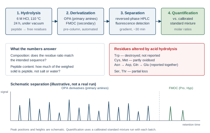
The peptide bonds have to be cleaved before the individual residues can be separated, and the analysis can only be as good as that step.4 The long-established condition is 6 M hydrochloric acid at 110 °C for 24 hours, with the sample held under vacuum or an inert atmosphere to limit oxidation.4
These conditions are a compromise rather than an optimum. Peptide bonds differ in how readily they are cleaved, amino acids differ in how well they survive the acid, and no single set of conditions both cleaves every bond completely and leaves every residue intact.4,5 Bonds between β-branched residues such as valine and isoleucine are cleaved slowly, so their yield can be low at 24 hours, while serine and threonine are progressively lost the longer hydrolysis continues.4,5 Where a particular residue is the point of the analysis, a hydrolysis time course can be run so the value is read from the trend rather than from a single time point.
Most amino acids have no useful chromophore or fluorophore of their own, so they are either detected after reaction with a reagent or converted to a derivative before separation.6 The classical instrument separated the free amino acids by ion-exchange chromatography and reacted them with ninhydrin after the column.1 Current practice more often derivatizes before the column and separates the derivatives by reversed-phase HPLC; the pharmacopoeial chapter describes both approaches alongside several reagent choices.6
LifeTein uses the pre-column OPA and FMOC combination. o-Phthalaldehyde reacts with a primary amine in the presence of a thiol to give a strongly fluorescent isoindole, which allows detection at low levels.2 That reaction needs a primary amine, so proline and hydroxyproline, whose amine is secondary, do not form it; 9-fluorenylmethyl chloroformate derivatizes those residues in the same automated sequence, and the two reagents together cover the full set.6 A further reagent, 6-aminoquinolyl-N-hydroxysuccinimidyl carbamate, derivatizes primary and secondary amines in one step and is an equally accepted alternative.3,6
Peak areas are compared with a standard mixture of known concentration run with the batch. The result is reported as the molar quantity of each residue recovered, from which the molar ratios and the peptide content are calculated.
Amino acid analysis and mass spectrometry answer different questions. A mass spectrum confirms that material of the expected mass is present and is the faster check on identity. It does not establish how much of the weighed solid is peptide, because it is not a quantitative measurement of mass against a standard. Amino acid analysis supplies that quantity. The two are complementary, and for a peptide going into a quantitative assay both are worth having.
Several residues do not survive the hydrolysis unchanged, and a report that did not say so would be misleading. These are properties of the method, not faults in a particular sample:4,5
| Residue | What happens during hydrolysis | Consequence for the report |
|---|---|---|
| Tryptophan | Largely destroyed by hot acid | Not reported from a standard hydrolysis; a separate alkaline or protected hydrolysis is required if the value is needed |
| Asparagine, glutamine | Side-chain amide is hydrolysed to the acid | Reported together with aspartate and glutamate, as Asx and Glx |
| Cysteine, methionine | Susceptible to oxidation | Recovery can be low and variable; oxidation to the stable derivative before hydrolysis gives a more reliable figure |
| Serine, threonine | Progressively degraded as hydrolysis continues | Values run low; a time course allows extrapolation back to zero time |
| Valine, isoleucine | Bonds between β-branched residues cleave slowly | Values can run low at 24 h; longer hydrolysis improves release |
Because of this, the residues used to anchor a quantitative result are normally the stable ones, and a composition is assessed on the pattern as a whole rather than on any single residue.
Amino acid analysis is a separately quoted analysis rather than part of a standard synthesis order, as is peptide content determination. Request it on the online quotation form or email sales@lifetein.com, and tell us the sequence and what the number is for — a composition check and a peptide content figure are quoted differently, and knowing which residues matter lets us choose the hydrolysis conditions sensibly.