Peptides are suitable for targeting protein-protein interactions and are used as smart delivery systems for targeting cancer cells. The main drawbacks of peptides are their susceptibility to enzymatic degradation and rapid kidney clearance. So, it is crucial to stabilize the peptides with unnatural modifications such as modifications of the peptide backbone, lipidation, attaching polymers, or modulating the primary, secondary, tertiary, or quaternary structure.
The peptide backbone can be modified to facilitate the peptide synthesis, increasing their solubility, and improving the pharmacological and pharmacokinetic properties. Many building blocks such as pseudoproline dipeptides are efficient tools for disrupting secondary structures. In peptide chemistry, some common practices are used for decreasing the fragility of peptides such as D-amino acids, azapeptides, methylation, backbone cyclization, or secondary structure constraints. This unnatural modification has a significant impact on the extended response and stability of peptides.
Lipidation using Cholesterol, fatty acid, or palmitic acid is a modification used for transforming peptides into peptide therapeutics. Lipids conjugated to peptides will increase their half-life by stabilizing their structure or binding to the cell membrane. The most prominent lipidated peptide drugs are insulin derivatives and glucagon-like peptide-1 (GLP-1) receptor agonists: liraglutide (Victoza). The combination of PEGylation and cholesterylation has been used for developing anticancer delivery systems to increase the antiviral potency of peptides and to boost their half-life in vivo.
The introduction of differently-sized polymers such as amphiphilic polyethylene glycol (PEG) is a highly popular tool in peptide chemistry. Each oxygen atom in a PEG polymer can bind two to three molecules of water, thus significantly increasing the size and solubility of the peptide attached to it. The PEGylated peptides have increased peptide solubility, lower immune response, and increased peptide bioavailability. There are fifteen PEGylated peptide drugs approved by the FDA.
How long do peptides last in the fridge? How should peptides be stored?
For long-term storage, the peptide should be kept in solid form in the deep freezer at < -15 °C. If stored at room temperature some peptides containing methionine or cysteine may begin to degrade. Therefore, we recommend storing them at -20C as soon as possible after receiving the package. At -20 or -80, the peptides will remain potent for 6 months or years before beginning to degrade. For short-time storage, a refrigerator (+4 °C) will suffice. Peptides should be protected from intense sunlight. Peptides containing fluorophores should be kept in the dark.
What may happen during the peptide storage?
Some typical degradation reactions or racemization may include the oxidation of Met, Trp, Tyr, or Cys. The deamidation of Asn, Gln, and the C-terminal amide may happen. The aspartimide may form. There might be the cleavage of Asn-Pro. The N-terminal Gln may form pyroglutamine. The dimerization of Trp and Tyr may form.
Are peptides soluble in water?
This is a list of non-polar, hydrophobic, or water-fearing amino acids: Ala (A), Ile (I), Leu (L), Met (M), Phe (F), Pro (P), Trp (W), Val (V).
Peptides containing 50% and more hydrophobic or nonpolar residues as above might be insoluble or only partly soluble in aqueous solutions. In this case, dissolve the hydrophobic peptides in a small volume of 50% (v/v) DMSO, DMF or acetonitrile in water first. Then add water or buffer to the desired concentration.
What is the best way to keep synthetic peptides in solution?
Dissolved peptides are less stable than the lyophilized dry powder. The solutions should be aliquoted before freezing to avoid thawing-refreezing cycles. Peptides containing Asn, Gln, Cys, Met, Trp, Tyr may be less stable because of the possible oxidization. The stock solutions should be in dry organic solvents to avoid premature hydrolysis. The buffer of pH 5-7 is considered as optimum for the stability of the aliquots. The peptide (>95% HPLC purity) solutions with cell cultures are frequently used without any sterilization. If the experiments are typically as short as a few hours, bacterial contamination wouldn’t be a problem.
Why do the dissolved peptides lose their activities? How to prevent peptides from losing their bioactivities?
The oxidation of Methionine yielding the sulfoxide might be the main reason for losing the peptide activity. The rate of sulfoxide formation is sequence-dependent. The best way is to replace the Methionine with its stable isostere Nle. Sulfotyrosine-containing peptides may lose their activity due to the desulfation.
Are peptides containing free Cys supplied as monomers? Will the Cys-containing Peptides be easily oxidized?
LifeTein provides Cys containing peptides as monomers unless specified otherwise. As air oxidation cannot be completely prevented, reducing the peptide before use by treatment with dithiothreitol (DTT) may be helpful to your experiment.
How to dissolve peptides containing disulfide bridges? How do I know if a cysteine in a peptide is free or oxidized?
Basic buffers should be avoided for peptides containing disulfide bridges. Peptides containing free thiol group may oxidize to form dimers or oligomers during storage, even as the lyophilized dry powder at a low temperature. Peptides provided as acetate salt are more sensitive to Cysteine oxidation than the corresponding TFA salt or HCL salt. The disulfide bond formation is rapid at neutral or slightly basic pH. Disulfide bridge formation is reversible. The disulfide bonds can be reduced at basic conditions using DTT. The pH 7-9.5 is the optimum pH range for reductions with DTT. The DTT solutions should be freshly prepared because DTT is readily oxidized.
How to dissolve peptides containing free cysteines?
Peptides containing a single free cysteine will be oxidized at pH>7 to form dimers. This oxidation can be reverted. Peptides containing two or more thiol moieties may yield a mixture of products upon oxidation. The pH 7.5-8 is the best condition for the disulfide bond formation. Hence, peptides containing free cysteines are best dissolved in degassed solvents, e.g. buffers pH<7, diluted acetic acid, 0.1% trifluoroacetic acid in aqueous acetonitrile. DMSO should be avoided, especially with peptide trifluoroacetates.
How to dissolve amyloid beta-amyloid peptide (1-42)?
The amyloid peptide Aβ (1-42), or other amyloid mutants may form insoluble aggregates during the storage. Aβ (1-42) is soluble in hexafluoroisopropanol (HFIP), DMSO, 0.1% aqueous ammonia, 50 mM TRIS ∙ HCl, or 1 mM NaOH. Reconstitution in HFIP or DMSO takes time whereas ammonia rapidly dissolves the peptide. The volatile solvent HFIP is usually evaporated leaving a residue of monomeric, soluble Aβ (1-42), which can be reconstituted with the chosen buffer at pH 7.4 to induce fibrillation. Aβ (1-42) solutions in DMSO or aqueous bases can be diluted directly with a working buffer.
How to dissolve amyloid beta protein (1-40)?
The freshly made Aβ (25-35) and (1-40) are soluble in oxygen-free water. Some old peptides may require the addition of acetic acid for dissolution. Do not dissolve the amyloid peptides in PBS. It is better to try water or 50% acetic acid and then dilute with the working buffer.
Is dry ice required for the shipment of peptides?
Dry ice is not required for the shipment of peptides. The lyophilizates in vacuum-sealed vials are sufficiently stable to tolerate 3-5 days at ambient temperature. Upon receipt, the product should be stored at the recommended storage temperature of -20 or -80.
Can I convert a free peptide or peptide with free acid into the peptide amide?
Unfortunately, the C-terminal carboxylic group cannot be converted to a carboxamide once the peptide is synthesized. The peptide amide has to be synthesized and cleaved in a different method.
Do I expect batch-to-batch variability?
The impurity profile and especially the peptide content can vary from batch to batch, even if we use the same standard protocols of synthesis and purification.
What is the difference between research-grade and GMP-API?
The Research-grade peptides are for laboratory and research purposes only! They may not be used as drugs for humans. The GMP-APIs are peptide products applicable to humans.
Why are peptides sold as salts?
Most peptides contain the basic functionalities: the guanidino group of Arg, the ε-amino group of Lys, the free N-terminus, and the imidazole moiety of His. These basic functionalities can form salts with acids. All our peptides are provided as trifluoroacetate salts unless specified otherwise. During cleavage from the carrier resin and purification, the peptide will react to the trifluoroacetic acid (a strong acid).
Additional ion-exchange steps are needed to make the acetate salt or HCL salt form peptides. Some acidic peptides, containing Asp, Glu, phosphor group, or sulfotyrosine, can form salts with bases and may be provided as ammonium salts.
How do you purify the peptides? How pure are your peptides?
Peptides obtained by SPPS are usually purified by the preparative RP-HPLC. The HPLC purity of most of our peptides is 95% or above. The lower purities may be expected for peptides containing free Cys, an N-terminal Gln, or sulfated peptides. The purity could be improved with the optimized synthetic and purification methods.
How do you synthesize small peptides?
Dipeptides, tripeptides, and other short peptides are usually synthesized in the solution called Liquid Phase Synthesis.
How do you synthesize peptides?
Fmoc- SPPS (solid-phase peptide synthesis) is the main method. The peptide is elongated starting from the C-terminus to the N-terminus of the sequence. An inert, insoluble but swellable polymer resin is used and the amino acids are added in the C→ N direction. After each coupling step, the Nα-blocking group is removed through repetitive protection and deprotection steps. LifeTein’s peptide synthesis method is fully automated.
How do you denote amino acids?
H- stands for an N-terminal free amino moiety, -OH for the unmodified C-terminal carboxyl group. H-Hyp-OH stands for – L-cis-hydroxyproline; H-Nle-OH is – L-norleucine;
Pyroglutamyl is abbreviated as Pyr. The natural peptides consist of L-amino acids. The D-enantiomer and the racemate are designated by -D- and -DL-, respectively.
What do L- and D-enantiomers mean?
All α-amino acids are asymmetric molecules except glycine. They can be obtained in two forms called enantiomers. Enantiomers differ considerably in their biological activity.
The D-enantiomer (D: dexter) represents the mirror image of the L-enantiomer (L: laevus). D- and L-enantiomers do not differ in their physical properties. Most L-amino acids rotate the light counterclockwise.
How to dissolve the peptide?
Families of Amino Acids:
Basic: Arg (R), His (H), Lys (K)
Non-polar (hydrophobic): Ala (A), Ile (I), Leu (L), Met (M), Phe (F), Pro (P), Trp (W), Val (V)
The solubility of a peptide is primarily dependent on the physical properties of its amino acids.
Basic peptides: The number of basic amino acids including the N-terminal amino group is more than the number of acidic amino acids including the C-terminal carboxyl moiety. The basic peptides may be dissolved in a small amount of an acidic solvent such as acetic acid or trifluoroacetic acid (TFA) and then diluted to the desired concentration.
Acidic peptides: The number of acidic amino acids including the C-terminal carboxyl group is more than the number of basic amino acids including the N-terminal amino group. The acidic peptide may be reconstituted in a small amount of a basic solvent such as 0.1% aqueous NH3 and then diluted with water to the desired concentration. The peptides containing free cysteines should be dissolved in the degassed acidic buffers because the thiol moieties could be rapidly oxidized at pH > 7 to disulfide bonds.
Neutral or highly hydrophobic peptides containing a high proportion of polar uncharged amino acids and/or hydrophobic amino acids should be dissolved in a small amount of an organic solvent such as DMSO, DMF, acetic acid, acetonitrile, methanol, propanol, or isopropanol and then diluted with water (or buffer) to the desired concentration.
Denaturating agents, such as urea or guanidinium hydrochloride may be used to solubilize peptides which tend to aggregate. The reconstitution of the aggregated peptide may take up to several hours. Sonication for several minutes in a water bath may be helpful to accelerate the dissolution of larger particles. The peptides containing Trp, Met, or Cys residues require special care to avoid oxidation.
What is peptide folding? Why do polypeptide chains fold?
Peptide folding is the process by which a peptide structure assumes its functional shape or conformation. By coiling and folding into a specific three-dimensional shape, the peptides can perform their biological function. The amino acids with hydrophobic side chains tend to end up clustered at the core of the structure so that they are out of contact with water. Covalent disulfide bridges can affect the shape of a protein. Protein folding is a spontaneous process because the Gibbs free energy is negative.
What are the four levels of protein folding?
There are four stages of peptide or protein folding, primary, secondary, tertiary, and quaternary. The secondary structure is the protein beginning to fold up. It can have two types of structure: the alpha helix, a coil shape held by hydrogen bonds in the same direction as the coil.
Why does peptide unfold at high temperatures?
Heat can be used to disrupt hydrogen bonds and non-polar hydrophobic interactions. This occurs because heat increases the kinetic energy and causes the molecules to vibrate so rapidly and violently that the bonds are disrupted.
What factors can disrupt the peptide structure and thus denature the peptide or protein?
Changing in pH and salt concentration will alter electrostatic interactions between charged amino acids. Higher temperatures reduce the strength of hydrogen bonds.
The presence of the reducing agents will break disulfide bonds between cysteines.
However, none of these agents breaks peptide bonds, so the primary structure of a protein remains intact when it is denatured.
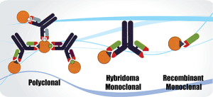
All forms of antibodies, polyclonal, hybridoma-based monoclonal, and recombinant monoclonal have both pros and cons as research tools.
The polyclonal antibodies display multi-epitope binding properties. However, the polyclonal antibodies could present variations in antibody performance. The monoclonal exhibit precise and reproducible binding properties from batch to batch. The recombinant monoclonal offers specificity and reproducibility and do not suffer from clonal drift or gene deletion. However, monoclonal antibodies are costly to produce.
Multi-epitope binding. The polyclonal antibody is an invaluable research tool if the antibodies are developed, and validated with the correct specifications. The polyclonal antibodies allow the binding of multiple antigenic determinants of the target. This enables polyclonal antibodies to be more sensitive with more excellent stability. The various binding of several different epitopes will make it more successfully bind a specific antigen in different immunoassays. For example, it is more effective at detecting a target for chromatin immunoprecipitation. The polyclonal antibody is a better option for many test conditions because their multi-epitope binding allows for antigen recognition even if some epitopes are affected by changes in an antigen’s tertiary structure or accessibility. However, the monoclonal antibody may have its epitope buried. Due to multi-epitope binding, it is more visible to use the polyclonal antibody for the western blotting experiment.
High stability. The polyclonal antibody is a mixture of antibodies with different biophysical attributes, such as charge and hydrophobicity. They are more resistant to changes in temperature and pH when compared with monoclonal antibodies. Usually, it is required to have stabilizing agents to prevent aggregation, and precipitation and preserve antibody binding for the storage of monoclonal antibodies.
A wide range of host animals. The polyclonal antibody can be generated in a wide range of host animals including rabbits, rats, mice, chickens, ducks, goats, sheep, donkeys, or even horses. The applications are flexible such as the quantity of the antigen, the injection methods and sites, the choice of adjuvants, and the duration of immunization schedules. The large animals with greater total blood volume will produce large amounts of antibodies over time because of the increased serum yield and the animals’ longevity.
Disadvantages.
Limited supply. This can be solved if the protocol is closely followed to guarantee reproducibility.
Cross-reactivity. This can be eliminated by negative absorption during affinity purification on a column containing the immunogens. The peptide antibody is especially valuable because of the specific immunogens with known sequences. The phosphor-peptide antibody production will go through the double screening process. First, the antisera would go through the non-phosphor-peptide column to remove the unwanted impurities. The elution will then go through the phosphor-peptide column for specific binding. Lot-to-lot consistency can be effectively managed and controlled by comparing the existing lots to historical lots.
It is highly challenging to deliver cosmetic actives and drugs to the skin because of the stratum corneum (SC). The permeability of large hydrophilic peptides or compounds across the stratum corneum on the outer skin surface is extremely low because of the lipophilic properties of the uppermost skin layer.
A short synthetic peptide was found to facilitate efficient transdermal protein drug delivery through the intact skin. Besides, the transdermal-enhancing activity of the peptide was sequence-specific and dose-dependent. The peptide creates a temporary opening in the skin barrier to enable insulin or other drugs to reach systemic circulation. So peptides hold potential as drug delivery vehicles for cosmetic purposes because of their simplicity, biocompatibility, and multi-functionality.
The delivery system was applied to treat melisma. Treatment with a cream formulation containing the transdermal peptide co-administered with other agents resulted in measurable inhibition of melanin production and melanocyte apoptosis. After four weeks of application, patients demonstrated a significant lightening of facial hypomelanosis lesions and almost wholly restored dark lesions to normal skin color after 12 weeks. These innovative peptide technologies give the patients even, young, and healthy-looking skin. Its topical application induces restoration of the skin’s most important structural dermal components.
LifeTein is working on a safe cell penetration peptide for co-administration with another short cosmetic peptide. A few short peptide candidates have been proven to significantly and visibly improve the skin’s evenness and overall appearance, and reduce keratinocyte-induced activation of melanocytes. Together with the cell penetration peptides, the cosmetic peptide acts safely and as a result inhibits the skin pigmentation process, which is of major cosmetic concern.
Self-assembly peptide into fibrillar nanostructures
Most of the potential therapeutic peptides have low solubility, chemical instability or low stability against protease. So it is essential to modify and optimize the peptides to improve the peptide bioavailability.
One novel approach is to create a self-assembly, highly ordered, and stable nanostructure. For instance, many peptide hormones including glucagon are stored in the form of β-sheet rich amyloid-like fibrils via a hydrogen bond network in the secretory cell.
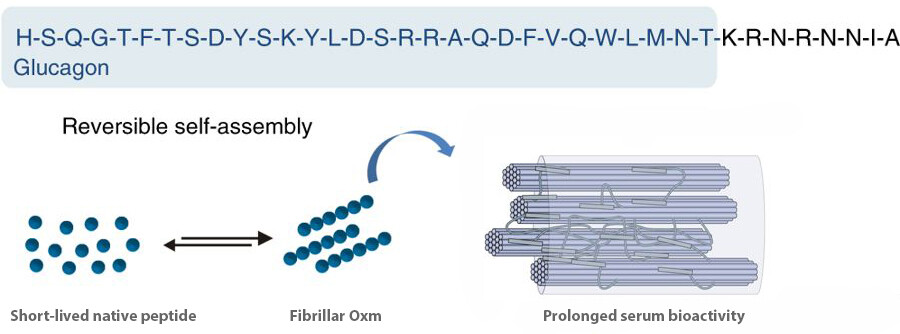
The oxyntomodulin is a peptide with a potential to treat obesity and diabetes. It is a 37-amino-acid proglucagon-derived peptide hormone with sequence homology to both glucagon and glucagon-like peptide-1 (GLP-1). The oxyntomodulin peptide self-assembles into a stable nanofibril formulation and later on releases an active peptide.
Here is the sequence of human oxyntomodulin: His-Ser-Gln-Gly-Thr-Phe-Thr-Ser-Asp-Tyr-Ser-Lys-Tyr-Leu-Asp-Ser-Arg-Arg-Ala-Gln-Asp-Phe-Val-Gln-Trp-Leu-Met-Asn-Thr-Lys-Arg-Asn-Arg-Asn-Asn-Ile-Ala, HSQGTFTSDY SKYLDSRRAQ DFVQWLMNTK RNRNNIA.
Oxyntomodulin self-assembles into fibrillar nanostructures
The peptide concentration was 10 mg/mL in water at an incubation pH of between 7.0 and 7.3 and low ionic strength (0.09% saline).
The solution was incubated at 37 °C and agitated by orbital shaking.
After incubation for five days, the solution turned turbid due to the formation of a suspension of aggregates. The conversion yield of the self-assembly of Oxyntomodulin into fibrillar nanostructures was estimated to be 99% under these conditions.
The nanofibrils were next used to seed a solution of free peptide at 10 mg/mL in water. 5. The solution was incubated without agitation for one week and then diluted to 1 mg/mL in 0.09% saline for another 2–9 days of incubation at 37 °C and then nine days at room temperature.
Free peptides were mainly in an α-helical conformation. The fibrillar Oxm showed the majority of β-sheet and some α-helical content.
Peptide nanofibrils dissociate to release intact peptide
1 mg/mL nanofibrils were incubated in water. 37% of the peptide was released after four hours of incubation.
In aqueous HCl, a 77% release was observed after only four hours. The peptide remained chemically intact after discharge from the nanofibrils
Benefits:
The released peptide is active and nontoxic in vitro.
The nanofibrils prolong peptide serum bioactivity in vivo. And there is no need to engineer or modify the original peptide. Other peptide hormones including glucagon, GLP-1, exendin-4, calcitonin, and gastric inhibitory peptide, are known to self-assemble. The self-assembly method may be used for the clinical application of reversibly self-assembling nanofibrils.
Reference: Controlling the bioactivity of a peptide hormone in vivo by reversible self-assembly. Nature Communications, volume 8, Article number: 1026 (2017)
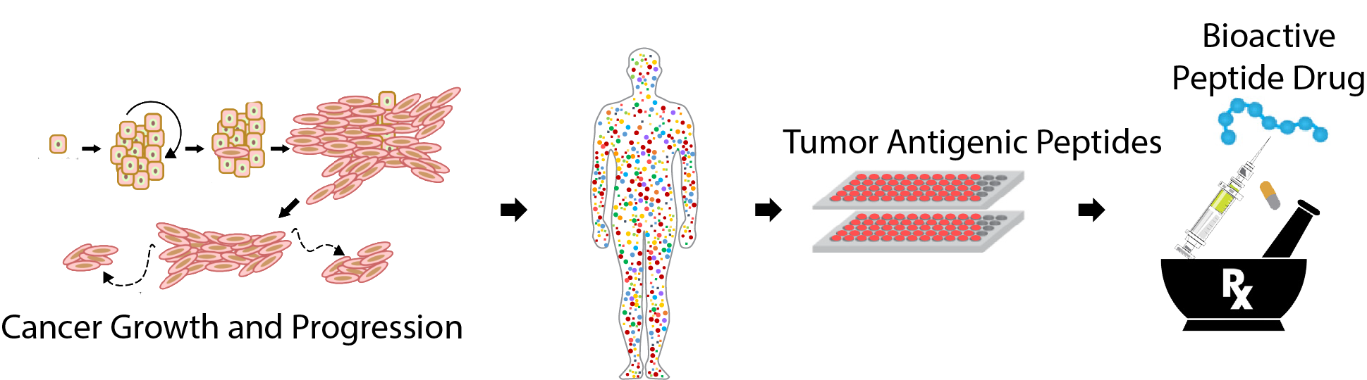
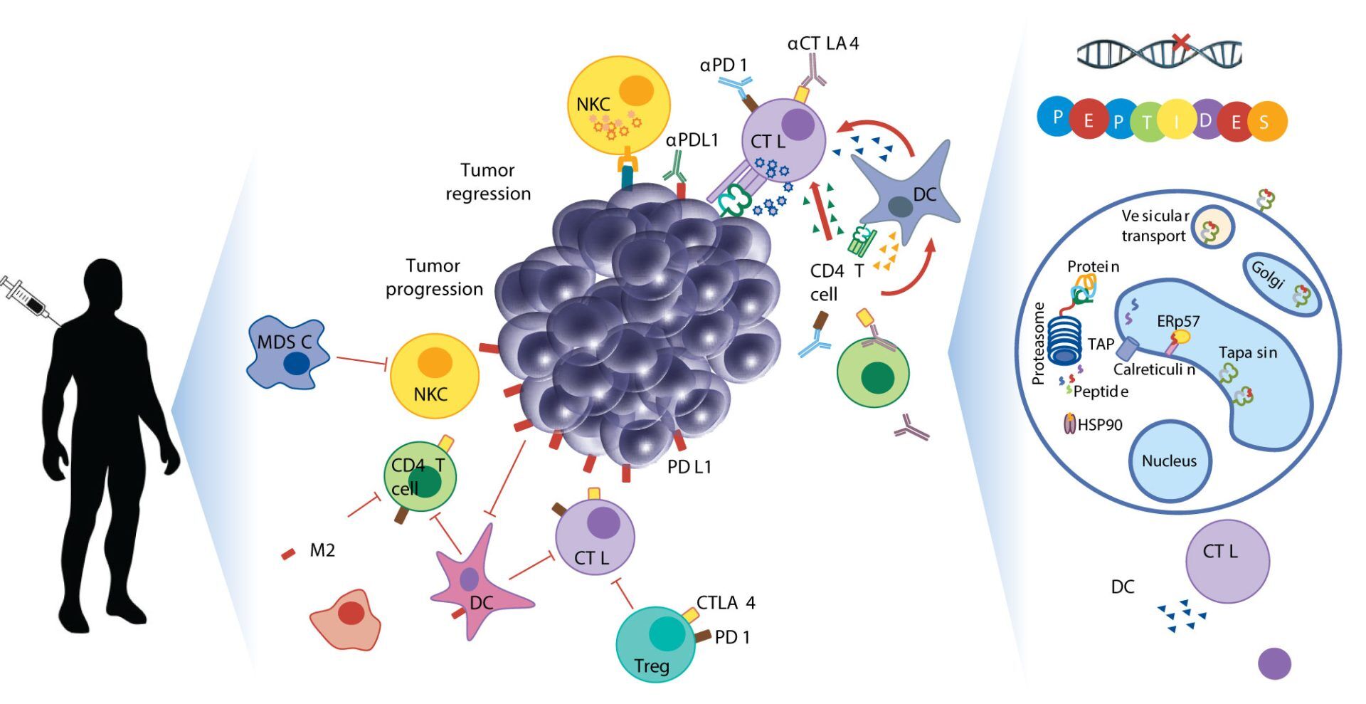
Cancer is a patient-specific disease, where no two tumors are alike. Neoepitopes are very frequent in all cancers. Amino acid substitutions can yield neoantigens that are detected by the immune system. So neoantigens have been used for therapeutic purposes such as identifying cancer variant peptides for diagnosis and treatment.
The neuropeptides have the following characteristics.
1. The 9-mer peptides are the most common among the high-binding neoantigens.
2. The neuropeptides have a hydropathy nature. The amino acid distributions, at all positions in neoepitopes of all lengths, contain more hydrophobic residues than the wild-type sequences.
3. Only a minority of predicted neoepitopes elicit protective tumor immunity. Peptide binding to a Human leukocyte antigen (HLA) molecule is a requirement for raising adaptive immunity.
How should we immunize against neoepitopes?
Since neoantigens are small peptides harboring tumor mutations, immunization with them usually needs strong immunostimulatory agents to produce an effective immune response.
Peptides as vaccines may not be able to stimulate the immune system powerfully enough on their own. Therefore, it is usually required to use an adjuvant in combination to elicit an effective immune response.
However, the MAPs-4 system, in which four copies of the same peptide epitope are synthesized on a lysine-based core, does not require a carrier protein, as the dense packing of multiple copies of an epitope in combination with a high-molar ratio produces a robust immunological response.
Accurate identification of neoepitopes and their subsequent use in cancer therapy is still in its nascent stages. With recent advances in Mass Spectrometry, faster and more precise identification of all expressed neoepitopes may be possible soon.
Neoepitopes as cancer immunotherapy targets: key challenges and opportunities
Protein-protein interactions (PPI) are highly specific electrostatic attractions between protein structures. The interactions regulate cell function and influence physiology and development. Mass spectrometry is often used to detect protein-protein interactions. However, all proteomic screens are differential and require two samples such as IP and mock IP, POI and wildtype, or POI and knockout.
The peptide/ biotin-peptide or peptide/fluorescence-labeled peptide are excellent tools for studying protein-protein interactions. Check this link for details: Protein-Protein Interactions: Methods for Detection and Analysis. https://www.lifetein.com/Peptide_Modifications_biotinylation.html
The compounds that can modulate PPI are hard to discover because the proteins have multiple binding sites and the screening assays are not reliable. In many cases, two or more proteins may interact with one another and form a complex. The optical fluorescence-based methods such as the Cy5, Cy7, FAM, FITC, TAMRA-labeled peptides, or FRET assay are particularly useful in these circumstances. Click for more details: https://www.lifetein.com/Peptide-Synthesis-FITC-modification.html.
The interactions between a fluorescently labeled or intrinsically fluorescent sample and a binding patterner are measured during the application. The changes in intrinsic fluorescence from tryptophan and tyrosine residues in the protein can be measured, which indicates transitions in the protein’s folding state.
The scientists have been working on fusion-based bifunctional proteins in cancer immunotherapy. The bifunctional protein sent an apoptotic signal to the tumor cells and enhanced their killing. The click chemistry is the perfect tool for the drug-protein or protein-protein conjugation. The more we understand the natural receptor-ligand complex and how it might signal, the better we can guide the design of therapeutic agonists. Click here for the peptide conjugation details: https://www.lifetein.com/price_modification_labeling.html
Epitope mapping identifies antigen regions that serve as binding sites for antibodies. The overlapping linear peptides derived from the primary sequence of the antigen are frequently used for epitope screening. Individual peptides can be divided into several fragments that overlap. The resulting overlapping peptide libraries can then be used for processes including continuous and linear epitope mapping.
For example, to map the epitope of an antibody, a few overlapping fragments spanning the target regions are constructed in an expression vector. These constructs are transiently transfected in cells and whole cell lysates collected after 48 hours are subjected to Western blotting with the antibody. To further map the region of this fragment, a series of overlapping peptides are synthesized. These peptides and lysates from the cells expressing the target full-length gene or empty vector (negative) are performed by the dot blot.
Mapping epitopes quickly and accurately are challenging because the epitopes tend to be nonlinear on antigens. Combining binding specifically toward two distinct epitopes into a single molecule can significantly enhance the immunotherapeutic properties of monoclonal antibodies. Multivalent interactions are the most efficient at driving IgE receptor signaling pathways.
There are several useful tools for studying antigen-antibody interactions.
Use the MAPs as the immunogens. Multiple Antigenic Peptides (MAPs) are peptides that are branched artificially, in which Lys residues are used as the scaffolding core to support the formation of 8 branches with varying or the same peptide sequences. For example, one goal is to include different epitopes from different virus proteins in a single unit. The epitopes showing the right prediction of antigenicity and conserved in most serotypes of a virus are selected and assembled as the MAP.
Screening combinatorial peptide libraries to optimize enzyme substrates and create high-affinity protein ligands. A critical biological application of custom peptide libraries is the characterization of the binding events that occur between specific proteins and their peptide ligands. A series of Overlapping Peptide, Truncation Peptide, Alanine Peptide Scanning, Scrambled Peptide, or Positional Peptide can be used for mapping and validating epitopes, the characterization of therapeutic antibodies, studying anti-antibody and neutralizing antibody actions in vitro.
Tat, the transcription activator of the human immunodeficiency virus type 1 (HIV-1) viral genome, enters cells in a non-toxic and highly efficient manner. Tat is the first known cell-penetrating peptide.
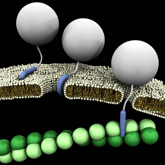
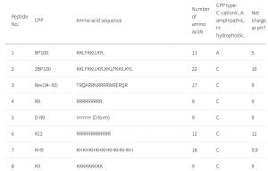
CPPs have been used as a carrier to deliver proteins or genes into cells and tissues. In this study, a CPP library composed of 55 CPPs was used to deliver genes into plant cells. Many CPPs showed efficient penetration into plant cells. The Lys-containing CPPs have higher penetration efficiency in the plant than in animal cells. This could be due to differences in lipid composition and surface charge of the cell membranes. No correlations were detected between the penetration efficiency and the cationic, amphipathic, or hydrophobic properties of peptides.
D-R9 is composed of D-form amino acids. D-R9 bound preferentially to the membrane and did not penetrate the cytosol or vacuole. In mammalian cells, poly-lysine-based CPPs are efficient and interact with membrane lipid head groups to induce wrapping of the membrane monolayers. Arg-rich peptides, such as the Tat peptide, are among the most efficient CPPs. Arg-rich CPPs may generate negative Gaussian membrane curvature to form pores or protrusions from endocytosis. The cell penetration efficiency of CPPs containing poly-Arg is higher than those containing poly-Lys. However, in a plant, Arg-rich CPPs are not the most efficient at penetrating plant cells.
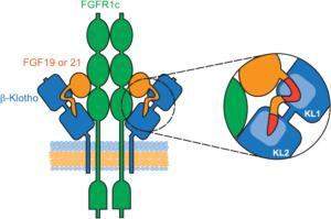
Endocrine fibroblast growth factors (FGFs) require Klotho transmembrane proteins as co-receptors to activate FGF receptor (FGFR) signaling.
A series of peptides were synthesized by LifeTein and used for the competition binding assay. Both the KL1 and KL2 domains of β-Klotho participate in ligand interaction. The FGF19 peptide was used for alanine scanning mutagenesis. It was found that a single amino acid mutation in either region was sufficient to abolish β-Klotho binding. FGF19 and FGF21 function through β-Klotho to regulate glucose and lipid metabolism.
How to perform the solid-phase binding assay
1. The 96-well plates were coated overnight at 4 °C with 2 µg/mL of antibody in PBS.
2. Plates were washed twice with PBST and blocked with 3% (w/v) BSA in PBS for 1.5 hours at room temperature.
3. The conditioned media containing β-Klotho were added to the plates and incubated for 1.5 hours at room temperature.
4. Plates were washed a few times.
5. The peptide mutation FGF21 and an anti-β-Klotho antibody were biotinylated with EZ-Link Sulfo-NHS-LC-Biotin at the indicated concentrations.
6. After washing, streptavidin-HRP was used for detection.
7. EC50 values were determined.
How to do a competition binding assay?
1. The WT and mutant peptides were custom synthesized and purified (>95% purity) by LifeTein.
2. Binding of FGF19 and FGF21 peptides to β-Klotho was assessed.
3. The β-Klotho ECD 6 × His, varying amounts of FGF19 and 21 peptides, and biotinylated human FGF19 or FGF21 protein were prepared.
4. The streptavidin donor beads and nickel chelate acceptor beads were added to the plates.
5. Plates were incubated for 3 hours at room temperature, protected from light, and read on the Plate Reader.
To provide the best experiences, we use technologies like cookies to store and/or access device information. Consenting to these technologies will allow us to process data such as browsing behavior or unique IDs on this site. Not consenting or withdrawing consent, may adversely affect certain features and functions.
Functional
Always active
The technical storage or access is strictly necessary for the legitimate purpose of enabling the use of a specific service explicitly requested by the subscriber or user, or for the sole purpose of carrying out the transmission of a communication over an electronic communications network.
Preferences
The technical storage or access is necessary for the legitimate purpose of storing preferences that are not requested by the subscriber or user.
Statistics
The technical storage or access that is used exclusively for statistical purposes.The technical storage or access that is used exclusively for anonymous statistical purposes. Without a subpoena, voluntary compliance on the part of your Internet Service Provider, or additional records from a third party, information stored or retrieved for this purpose alone cannot usually be used to identify you.
Marketing
The technical storage or access is required to create user profiles to send advertising, or to track the user on a website or across several websites for similar marketing purposes.