The Power of Nucleic Acid Mimics (NAMs) in Advanced Genetic Research

Comparison of the most important properties of DNA and NAMs probes (a) and PNA and LNA probes (b).

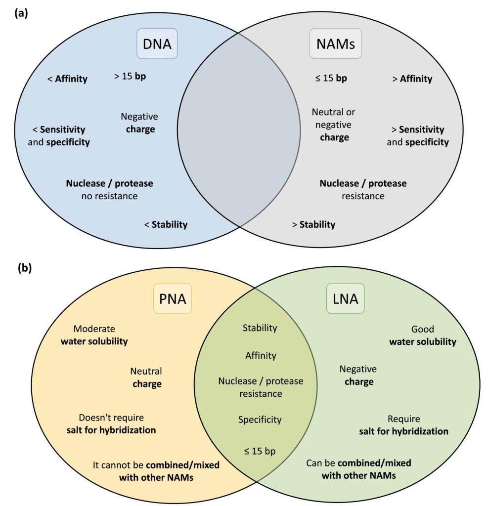
The landscape of in situ hybridization (ISH), specifically fluorescence in situ hybridization (FISH), has undergone a transformative shift with the introduction of Nucleic Acid Mimics (NAMs). These modified probes, encompassing Peptide Nucleic Acid (PNA), Locked Nucleic Acid (LNA), 2′-O-Methyl-RNA, UNA (unlocked nucleic acid), and Phosphorodiamidate Morpholino Oligomers (PMOs), have emerged as groundbreaking tools, overcoming the limitations associated with traditional DNA and RNA probes.

Peptide Nucleic Acids (PNAs):

Peptide Nucleic Acids (PNAs) stand at the forefront of this molecular revolution, offering a fusion of DNA specificity and peptide versatility. The distinctive PNA backbone, composed of peptide linkages, ensures unparalleled stability and resistance to enzymatic degradation. With superior hybridization properties, PNAs bind to complementary DNA or RNA sequences with exceptional affinity, making them indispensable for applications ranging from targeted gene therapy to diagnostic assays and antisense technologies.

Unlocking the Potential: PNA’s Key Features:

Stability and Resistance:
PNAs, characterized by a neutral polyamide backbone, showcase remarkable stability against nucleases and enzymatic degradation. This attribute enhances the half-life of PNA molecules, ensuring their efficacy in diverse experimental conditions.

High-Affinity Binding:
The hybridization capabilities of PNAs are unparalleled, facilitating strong, sequence-specific binding to target nucleic acids. PNA’s shorter length allows for enhanced cell penetration and consistent hybridization performance, even under low salt concentrations. Its unique melting temperature response to single nucleotide changes enables precise probe design.

Versatility in Applications:
PNAs find applications across a spectrum of research areas, including molecular diagnostics, gene editing, and nanotechnology. Their adaptability for specific sequences and functions makes PNAs an invaluable asset in the molecular biologist’s toolkit.

Locked Nucleic Acid (LNA):

Described in 1997, Locked Nucleic Acid (LNA) boasts a ribose ring locked in a specific conformation, ensuring water solubility and low toxicity. LNA’s design flexibility, including modifications like phosphorothioate, enhances resistance to nucleases without compromising affinity. Combining LNA with 2′-O-Methyl-RNA provides flexibility in melting temperature adjustments for optimized hybridization efficiency.

UNA and Other NAMs:

UNA, an acyclic RNA analog, offers flexibility, though it may impact nucleic acid duplex stability. Modifications like 2′-pyrene and 3′-O-amino-UNA address stability concerns, presenting potential applications in FISH experiments. Additionally, Phosphorodiamidate Morpholino Oligomers (PMOs), characterized by non-ionic properties and resistance to nucleases, have shown success in bacterial and fungal infection detection via FISH.

Challenges and Progress:

Despite the exceptional qualities of NAMs, particularly PNA and LNA, their widespread adoption in FISH for microorganism detection has been slower than anticipated. Challenges include hydrophobicity and water solubility issues for PNA probes, along with a general lack of awareness among laboratories. Nevertheless, studies showcasing PNA’s superior performance over traditional DNA probes underscore the promising potential of NAMs in microbial detection.

Conclusion:

As research in the field progresses, ongoing efforts are addressing the limitations associated with NAMs. Their unique features position them as compelling alternatives for FISH-based microorganism detection, holding the promise of unlocking new frontiers in advanced genetic research.

A New Dawn in Ulcerative Colitis Care: Peptide-Assisted Antigen-Specific Immunotherapy

The chronic autoinflammatory bowel disease ulcerative colitis affects millions around the world. The condition involves autoreactive T cells and macrophages in the colonic mucosa attacking healthy colon cells, leading to inflammation, ulcers, and other debilitating symptoms and complications. While there is no outright cure, the only treatment involved is long-term immunosuppression, which can lead to even more health complications and risk of cancer down the line. One novel solution possible is antigen-specific immunotherapy, where the specific antigens are presented to the T cells in the presence of immunomodulators. Peptides assist antigen-specific immunotherapy of ulcerative colitis by being adsorbed to nanofibers utilized in the colon-specific niche developed for this condition.

Mimetic peptides utilized in antigen-specific immunotherapy

LifeTein provided the group with the Cationic TGF-β1 mimetic peptide, whose role in the niche is to bind to a key receptor and suppress the activation of CD8+ T cells. This also polarizes the macrophages involved as well. The final results proved that not only could the auto reactive T cells be inhibited, but healthy colon cells could help repair previous damages of ulcerative colitis afterwards. All of this was achieved while actively avoiding the cancer risks of standard immunosuppressive approaches. Hopefully, this modular method can be pivotal in future developments for safer and more effective uses of immunotherapies.

Kin Man Au, Justin E. Wilson, Jenny P.-Y. Ting, Andrew Z. Wang. An Injectable Subcutaneous Colon-Specific Immune Niche For The Treatment Of Ulcerative Colitis doi: https://doi.org/10.1101/2023.10.03.560652

Diving into Live-Cell Imaging: Prerequisites, Techniques, and Expert Insights

Live-cell imaging using peptides serves as an invaluable asset in the world of life science research, offering a window into the dynamic lives of cells in conditions closely resembling their natural habitat. By capturing real-time images of living cells, researchers can explore intricate aspects of cell behavior, growth, and movement.

In contrast to the static imaging of fixed cells and tissues, where photobleaching poses a significant challenge, live-cell imaging calls for a different approach. While fixed cell imaging demands high-intensity illumination and prolonged exposure, such conditions prove detrimental to the vitality of live cells. Consequently, live-cell microscopy finds itself in a constant balancing act, seeking to attain image quality while safeguarding cell well-being. This balance often imposes limitations on spatial and temporal resolutions in live-cell experiments.

Live-cell imaging encompasses a wide range of contrast-enhanced techniques in optical microscopy. The majority of investigations rely on various forms of fluorescence microscopy, often in conjunction with transmitted light methodologies. The ongoing evolution of imaging techniques and the development of fluorescent probes ensure that live-cell imaging retains its significance as a critical tool in the field of biology.

  1. The Fundamentals of Fluorescence Microscopy

One of the fundamental considerations is determining the precise amount of excitation light required to obtain a meaningful image. It’s worth noting that high-intensity light, especially in the near-UV range, can harm cells and potentially induce DNA damage. However, in live-cell fluorescence microscopy, the primary source of phototoxicity stems from the photobleaching of fluorophores. A critical protective measure involves deactivating the illuminating light when not in use. Employing shutters to control light exposure proves to be a pivotal element in live-cell imaging.

Additionally, it’s crucial to eliminate undesired wavelengths of light and opt for emission filters that are fine-tuned to maximize the signal. Mitigating photobleaching can be achieved by reducing oxygen levels, and minimizing background fluorescence can be accomplished by excluding phenol red and serum from the culture medium. It’s also essential to prevent any contamination of the illuminating light with even minute traces of ultraviolet or infrared wavelengths. The contained photobleaching chemistry within the β-barrel structure of fluorescent proteins (FPs) or peptides makes them less phototoxic.

The most effective strategy for reducing photobleaching and the associated photodamage is to minimize excitation light exposure by carefully managing exposure time and light intensity while maintaining a satisfactory signal-to-noise ratio tailored to the specific research question.

  1. The Live Cell Imaging Microscope

When selecting an optical microscopy system for live-cell imaging, three key factors come into play: detector sensitivity (signal-to-noise ratio), specimen viability, and the speed required for image acquisition. To optimize the signal-to-noise ratio, it’s crucial to select filters that closely match the spectral profiles of the fluorophores in use. Most epi-illumination microscopes and confocal systems acquire data in four dimensions.

Time-lapse imaging, involving the capture of cellular events over various timeframes, is commonly employed. This technique enables the repetitive imaging of cell cultures at specific time intervals. Wavelengths for excitation and emission filters should be finely tuned to match the fluorophore, thus reducing unnecessary light exposure. To minimize photodamage to the specimen, it’s advisable to use the lowest magnification that suits the specific experiment.

  1. Managing the Microscope Environment

Maintaining a physiological environment is imperative for cultured cells and tissues to behave naturally. Thus, the control of factors such as temperature and tissue culture medium composition plays a critical role in obtaining meaningful data in live-cell imaging experiments. Mammalian cell lines are typically maintained at 37°C. Variations in temperature and vibrations can negatively affect focus stability. To address these issues, options like stage-top incubators and fully enclosed microscopes can be considered. Most tissue culture media are buffered to a physiological pH using sodium bicarbonate and 5% CO2.

  1. Unmasking the Potential of Fluorescence Resonance Energy Transfer (FRET)

Fluorescence resonance energy transfer (FRET) is a remarkable technique that enhances the spatial resolution of fluorescence microscopes to under 10 nanometers. This substantial improvement in resolution makes FRET particularly appealing for studying co-localization events in biological samples, especially within living cells. However, in live-cell studies, the risk of FRET measurements being invalidated by acceptor fluorophore recovery, similar to FRAP experiments, must be carefully considered. Therefore, the use of acceptor photobleaching in live-cell experiments may not always be suitable.

Useful FRET calculator that provides a listing of key FRET pair information: www.fpbase.org/fret/

Furthermore, modern fluorescent dyes, such as the Alexa dye series, offer advantages like enhanced quantum efficiency and brightness, making them indispensable for specific techniques. LifeTein offers an extensive range of fluorescent labeling options, including FITC, FAM, TAMRA, Cyanine Dye Cy3, Cy3.5, Cy5, Cy5.5, Cy7, Cy7.5, EDANS/Dabcyl, MCA, AZDye, BODIPY FL or Alexa Fluor (Alexa488, Alexa532, Alexa546, Alexa594, Alexa633, Alexa647), ATTO Dyes (Atto465, Atto488, Atto495, Atto550, Atto647), and DyLight (DyLight 488, DyLight 550). However, chemical fluorescent dyes often exhibit higher cytotoxicity, possibly due to the cytoplasm’s reduced protection from reactive free radical breakdown products when compared to FPs, where the fluorophore is encapsulated within the FP beta-barrel structure. A range of cell-permeable fluorescent molecules is available for the specific labeling of intracellular organelles.

  1. Navigating Photobleaching

Photobleaching is an inherent aspect of live-cell imaging. Even the most advanced fluorescent molecules convert only a fraction of the absorbed energy into fluorescence. Some of this energy inevitably triggers chemical reactions that lead to fluorophore breakdown. Thus, aside from maintaining a physiological environment and confirming the specific labeling and function of the protein or organelle of interest, the most critical factor for successful live-cell imaging and obtaining meaningful data is the minimization of excitation light. This requires a thorough understanding of the microscope and optimization of components that regulate exposure, wavelength selection, and the collection of emitted photons.

A proper experimental setup is equally vital. Waiting for the visual system to adapt to darkness before attempting to locate a faint sample is advisable, given that modern cameras are often more sensitive than the human eye. However, it’s important to avoid using live camera modes to find your sample. Every photon is precious.

Unfortunately, photobleaching remains an unavoidable challenge, often determining the number of images that can be acquired. There is no simple solution to eliminate photobleaching completely. The specific excitation and emission bands, as well as the intracellular environment, influence the apparent brightness and, consequently, the apparent photobleaching of specific fluorescent proteins.”

Alzheimer’s disease and the role of β-amyloid

The incurable neurodegenerative Alzheimer’s disease has long been associated with β-amyloid build-up in the brain. While this has been known, direct evidence supporting the close relationship of Alzheimer’s disease and the role of β-amyloid has been hard to come by, until now. Recent Aβ-immunotherapy trials have shown that removing aggregated β-amyloid from symptomatic patients can slow down the disease.

β-amyloid removal slows the progression of Alzheimer’s disease

This breakthrough holds many implications for future treatment and handling of Alzheimer’s disease. While the new evidence is far from being a cure itself, it presents the opportunity for long-term prevention and potential immunoprevention towards the disease. This is in part due to how early the signs and abnormal β-amyloid build-up can begin in Alzheimer’s patients, as well as how complex the disease itself can become. However many clinical trials and experiments it may take, groups like LifeTein will always be ready to help supply researchers with the materials they need to make this future possible.

Jucker, M., & Walker, L. C. (2023). Alzheimer’s disease: From immunotherapy to immunoprevention. In Cell (Vol. 186, Issue 20, pp. 4260–4270). Elsevier BV. https://doi.org/10.1016/j.cell.2023.08.021

How to Design Cell Penetrating Peptides?

Please view this video on how to design cell-penetrating peptides. The transcript is listed below.

Transcript

Slide 1:

Thank you for joining me. My topic today is the cell-penetrating peptides. My main focus will be the peptide design, peptide synthesis, and its applications.

Slide 2:

First, let me briefly introduce LifeTein. LifeTein was founded in 2008. We have been in the peptide industry for more than ten years. We specialize in peptide synthesis, chemical synthesis, antibody production, and protein services.

Slide 3:

Our main focus is peptide synthesis service. However, over the years, we have expanded to other protein-related areas like protein, antibody services, and products.

Slide 4:

So let us quickly get into the topic: cell-penetrating peptides. What is a cell-penetrating peptide or cpp? From the definition, CPP is a short peptide. They can be about 4-40 aa. The short peptide can enter the cell membrane. They can deliver bioactive cargoes.

Slide 5:

CPPs can also be used to deliver bioactive cargos like siRNAs, DNA, polypeptides, liposomes, nanoparticles, and others, in cells for therapeutic or experimental purposes.

Slide 6:

There are a few popular models for CPP’s entry. 1. The inverted micelle model. The CPPs are positively charged. They interact with the negatively charged phospholipids in the membrane. 2. Direct entry or direct translocation. For example, sequences with multiple Arginines can cause a short-time membrane cytolysis and enter the cells directly. 3. By the traditional method of endocytosis. I will not talk about the details.

Slide 7:

Here are a few examples of the CPP. The most famous examples are HIV tat sequence. The TAT peptide is arginine-rich and can directly penetrate the plasma membrane and stabilize DNA.

Another example is the arginine-rich peptide R8 or R9. We can add stearic acid to the N-terminus.  Stearic acid is a saturated fatty acid with an 18-carbon chain. If you would like to do live cell imaging, we can add fluorescent dye such as Fitc, Alexa fluor, or Cy dye at the N-terminus or C-terminus. I will get to the details.

Slide 8:

CPPs can enter the regular cell membranes. Some other peptides are tissue-targeting peptides. For example, this brain-homing peptide can cross the blood-brain barrier. Other peptides can cross the skin as transdermal peptides, target heart tissues as cardiac targeting peptides, and nuclear localization signal peptides.

Slide 9:

On this slide, I will talk more about the peptide design. There are many ways to make the peptide permeable. In the case of DNA or RNA, you can simply mix the CPPs with oligos. Many transfection reagents are using this mechanism. Simply put, DNA is negatively charged and peptide is positively charged. If you mix them together, they will form small micelles for cell penetration.

However, most of our work is to put the CPPs and your target together by covalent links. For this example, we put eight arginines at the N-terminus of your peptide. A linker called Ahx is added as a spacer. Some users prefer no spacers. It seems that both worked for the purpose. The eight arginines can be put at the C-terminus as well. According to the feedback from our users, most of the N-terminal CPP worked well. A few worked well for the C-terminal conjugation. I guess it depends on the projects.

This example is the Npys linker modification. The cysteine is added to your peptide. We conjugate two sequences together to form a disulfide bond. This is especially useful for the cancer study. Cancer cells have a lower pH of 6.7-7.1. Normal cells have a higher pH of 7.4. Under the acidic environment, the disulfide bond can be cleaved. If your target peptide is a cancer drug candidate, the CPP can introduce the drug cargo to the cancer cell and release the target within the cell. These disulfide-based prodrugs are important for cancer therapy.

If the cysteine is not available for your case, we can add a compound called lysine azide. This method needs click chemistry.

Slide 10:

There are two kinds of click chemistry. The one with copper as the catalyst and the one without. The preferred method is copper-free click chemistry. It is called DBCO and azide reactions. The final conjugate will have a large linker. Many scientists have concerns about the bulky size of the linker. However, some drugs contain bulky linkers without issues or side effects.

Back to Slide 9:

Let us go back to the sequence. The design does not have to be this way. The lysine azide can be any place in the sequence. If you have a head-to-tail cyclic peptide, you can add the lysine azide in the middle. The final product will be like a lollipop, with the CPP as the tail. If the N-terminus is very important to you, you can add the azide at the C-terminus.

Slide 11:

Let us move on to other scenarios. If you would like to track the peptides in live cells, fluorescent dyes can be added. We can do Fitc, Fam, Cy3, cy5, Cy7 and Alexa Fluor. This design will give direct evidence that your target is inside the cells.  

There is a different kind of peptide called peptide nucleic acid or PNA. It is DNA or RNA analog. We can synthesize half as peptide and another half as the PNA.  

This structure is the one I just mentioned earlier. The cyclic tumor targeting RGD peptide can be linked with an R8 cell-penetrating peptide to form a lollipop-shaped structure.

Slide 12:

So far, we have mentioned different ways to conjugate the cargo with cell-penetrating peptides. If your targets are nanoparticles or gold particles. Our requirement is to have active groups like a thiol group or a free amine on it. We have to have the active groups react to the cell-penetrating peptides. It is the same requirement for small compounds.

Slide 13:

The last concept I would like to introduce is the antibody-drug conjugate. This concept is widely accepted in the antibody drug industry. There are three important components: an antibody, a cleavable linker, and the drug.  Once the antibody binds to the target, the drug is released after the hydrolysis by protease.

Slide 14:

The same concept can be used for the peptides. For this concept, we need to screen the best drug candidate for cell entry. The CPP can be tumor-homing peptides, brain-homing peptides, or cardiac targeting peptides I mentioned earlier.

Slide 15:

First, we need to modify the compound. It is better to have a free amine in the compound. Then we can modify the amine group to an azide group. Afterward, we can use the click chemistry for the following conjugation.

Slide 16

LifeTein produced a series of CPPs. They are ready to conjugate your compounds for screening. So far, we have designed and produced more than fifty CPPs.

Slide 17

Step 3 is conjugate peptides with drug candidates.

Slide 18

Once the CPP is conjugated with the drug compound using click chemistry, we can send the final product back to you for further screening. The purpose is to find the best drug delivery system.

Slide 19

To summarize today’s topic, I talked about cell-penetrating peptides with different cargos. As long as you have an active chemical group on the nanoparticles, compounds, or liposomes, we can conjugate the target to any peptide.

Slide 20

That is all for today. Please let me know if you have any questions. Please feel free to contact us by email or phone calls.   

Revolutionizing Peptide Synthesis: A Breakthrough Cocktail For Methionine-Containing Peptides

In the world of peptide synthesis, a game-changing innovation has emerged – a remarkable cocktail designed to enhance the cleavage and deprotection of methionine-containing peptides. This groundbreaking concoction, known as Reagent H, is set to transform the landscape of solid-phase peptide synthesis, particularly for those using the 9-fluorenylmethoxycarbonyl (Fmoc) methodology.

Unveiling Reagent H: Your Key to Methionine Side-Chain Protection

Reagent H, comprised of trifluoroacetic acid (81%), phenol (5%), thioanisole (5%), 1,2-ethanedithiol (2.5%), water (3%), dimethylsulphide (2%), and ammonium iodide (1.5% w/w), has been meticulously crafted to minimize the pesky oxidation of methionine side chains during synthesis. Its exceptional performance is exemplified in the synthesis of a model pentadecapeptide from the active site of DsbC, a pivotal player in protein disulfide bond formation.

The Triumph of Reagent H: Methionine Sulphoxide Conquered

When put to the test, Reagent H outshone its competitors, cocktails K, R, and B. The crude peptides obtained from these widely used mixtures contained a staggering 15% to 55% of methionine sulphoxide. However, Reagent H demonstrated its prowess by yielding pristine peptides devoid of methionine sulphoxide. Remarkably, even when 1.5% w/w NH4I was added to cocktails K, R, and B, they couldn’t match the perfection achieved by Reagent H, although their yield of the desired peptide fell short.

Unraveling the Mysteries: A Closer Look at Reagent H’s Mechanism

But how does Reagent H achieve this remarkable feat? We delve into the proposed mechanism behind its in situ oxidation of cysteine, shedding light on its impressive ability to safeguard methionine side chains while delivering high-quality peptides.

In the world of peptide synthesis, Reagent H stands as a beacon of hope for researchers seeking purity, precision, and protection in their work. Its ability to minimize methionine side-chain oxidation is nothing short of revolutionary, promising a brighter and more efficient future for peptide synthesis enthusiasts. Say goodbye to impurities and hello to perfection with Reagent H.

Peptides Help Determine the Effects of Aging on Vision

Many sensory declines come along with aging, one of much concern being sight. That being said, there is a need for much more research on the visual changes associated with such aging. Specifically, the changes of rod bipolar cells and their ribbon synapses due to aging are an area of interest, along with the complex calcium systems at work. Using a zebrafish model, peptides help determine the effects of aging on vision and the retina.

TAMRA-RBP peptides tag rod bipolar cells

Zebrafish were used for this experiment thanks to their unique roles as model organisms; they share 70% genomic similarity with humans, and their short lifespan offers the chance to study life cycles in a few short years while still comparable to human aging over decades. Researchers compared data between middle-aged (MA, 18-months-old) and older-aged (OA, 36-months-old) zebrafish, equating to human ages of approximately 38 and 75 years of age, respectively. Using TAMRA ribbon-binding peptides from LifeTein, the team was able to observe changes between the two ages of zebrafish.

What was discovered was a decreased number of synaptic ribbons and increased ribbon length in the OA models. Further, there were many alterations to the local calcium dynamics of the system, implying a more complex change to vision deterioration than initially expected. The model shows how subtle changes could have vast implications for disease models where these alterations may be amplified, and surely sheds more light on how human vision may decline with age.

Abhishek P Shrestha, Nirujan Rameshkumar, Johane Martins Boff, Rhea Rajmanna, Thadshayini Chandrasegaran, Frederick E Courtney, David Zenisek, Thirumalini Vaithianathan
bioRxiv 2023.09.01.555825; doi: https://doi.org/10.1101/2023.09.01.555825


Innovative Approach to Combat Breast Cancer: The Impact of F11R/JAM‑A Peptide Antagonist on Metastasis

LifeTein’s Innovative Peptide Antagonist: A New Ally in the Fight Against Breast Cancer Metastasis

Breast cancer stands as the most commonly diagnosed cancer in women worldwide and is a leading cause of cancer-related deaths. Among its subtypes, triple-negative breast cancer (TNBC) is particularly notorious for being aggressive and prone to metastasizing to vital organs like the brain, lungs, bones, and liver. Despite being more responsive to chemotherapy, TNBC’s propensity for metastasis poses a significant challenge in cancer treatment.

Recent studies, including notable research from the Medical University of Lodz in Poland, have identified a key factor in TNBC metastasis: increased F11R/JAM-A activity. This protein plays a crucial role in the early stages of cancer cell migration across blood vessels, a precursor to metastasis. Enter LifeTein, a pioneering force in peptide technology, which has made a groundbreaking contribution to this research area.

LifeTein provided a specialized peptide antagonist, named P4D, designed to specifically target and inhibit F11R/JAM‑A. The effectiveness of P4D was rigorously tested in lab models. Remarkably, this antagonist not only curbed the proliferation of TNBC cells but also significantly reduced their survival by directly targeting F11R/JAM-A. The result was a notable hindrance in the metastasis process in the mouse models used for the study.

This breakthrough has significant implications. The success of P4D in these preliminary studies suggests potential for future clinical trials and paves the way for more targeted, effective treatments for TNBC, possibly extending to the development of tailored antibodies. LifeTein’s contribution to this field exemplifies its commitment to advancing cancer therapy, offering new hope to those battling with TNBC.

For more detailed insights, refer to the original study by Bednarek, R., Wojkowska, D.W., Braun, M. et al., titled “Triple negative breast cancer metastasis is hindered by a peptide antagonist of F11R/JAM‑A protein,” published in Cancer Cell International.

Bednarek, R., Wojkowska, D.W., Braun, M. et al. Triple negative breast cancer metastasis is hindered by a peptide antagonist of F11R/JAM‑A protein. Cancer Cell Int 23, 160 (2023).

Innovative Peptide Technology in Click Chemistry: Paving the Way for Smarter Drug Delivery

As methods of medicine advance, targeted drug delivery becomes a more appealing and achievable option over its non-selective counterpart. It can focus solely on increasing therapeutic concentration in the target area while greatly eliminating any exposure to healthy tissue, and thus drastically lowering side effects as well. The effective and simple mechanisms of click chemistry are a great way to design payloads for these targeted drug delivery methods. With the use of enzyme-degradable peptides in click chemistry drug delivery, lasting therapeutics can remain in the system for local sustained release over time as well.

Enzyme-degradable peptides for sustained drug delivery

The team at Rutgers focused on a two-phase method to set up the targeted drug delivery. First, ROS-sensitive PEGDA and acrylate-PEG-azide are aimed at the target area, driven by elevated free radical levels. Once the pretargeting is complete, a payload tethered to DBCO is delivered and captured via azide-DBCO reactions. Enzyme-degradable peptides were provided by LifeTein and incorporated into both steps for the ongoing release of the captured payloads.

The results showed success in the models tested, with the initial dosage still effective in capturing the payload several days later. This system demonstrated the versatility of a two-phase method, where long-term effects are even further avoided by incorporating enzyme-degradable peptides. The proof of concept displayed here has great promise for the future of drug delivery and just goes to show how applicable click chemistry is to even more fields.

Emily T. DiMartini, Kelly Kyker-Snowman & David I. Shreiber (2023) A click chemistry-based, free radical-initiated delivery system for the capture and release of payloads, Drug Delivery, 30:1, DOI: 10.1080/10717544.2023.2232952

Convergent Evolution of Holocentromeres Studied with Peptides

During cell division, microtubules in the chromosome attach to a region called the centromere. While most species have a single size-restricted centromere, or a monocentromere, some species exist with multiple centromeres distributed across the chromosome, called holocentromeres. What is even more interesting is how holocentric chromosomes are considered to have evolved from the monocentric organisms, and this transition occurred independently across distant lineages, such as green algae, protozoans, invertebrates, as well as flowering plant families. One group aimed to study these holocentromeres more via the lilioid Chionographis japonica. Their goal was to better understand the convergent evolution of holocentromeres studied with peptides.

Peptides help explore holocentromeres

The group determined that the chromosomal localization of the target centromere is usually marked with histone H3 (CENH3). With this knowledge, they utilized peptides and antibodies of CENH3 provided by LifeTein to create models of the transition of C. japonica from interphase to prophase and study the possible mechanisms as well. They found the holocentromere was made up only of a few, evenly spaced CENH3-positive megabase-sized satellite arrays. Overall, the reason for the convergent evolution of holocentromeres from a monocentromere may stem from multiple factors, but more experiments like the ones presented will surely provide further analysis into this complex and fascinating case of convergent evolution.
Kuo, YT., Câmara, A.S., Schubert, V. et al. Holocentromeres can consist of merely a few megabase-sized satellite arrays. Nat Commun 14, 3502 (2023). https://doi.org/10.1038/s41467-023-38922-7